 Movie
Movie Controller


+ Open data

- Basic information
| Entry | Database: PDB / ID: 6orh | |||||||||
|---|---|---|---|---|---|---|---|---|---|---|
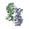
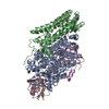
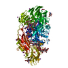
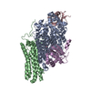
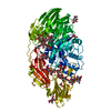
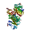
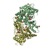
| Title | Crystal structure of SpGH29 | |||||||||
 Components Components | Glycoside hydrolase | |||||||||
 Keywords Keywords | HYDROLASE / glycoside hydrolase | |||||||||
| Function / homology |  Function and homology information Function and homology informationalpha-L-fucosidase / alpha-L-fucosidase activity / fucose metabolic process / glycoside catabolic process / lysosome / metal ion binding Similarity search - Function | |||||||||
| Biological species |  Streptococcus pneumoniae serotype 4 (bacteria) Streptococcus pneumoniae serotype 4 (bacteria) | |||||||||
| Method |  X-RAY DIFFRACTION / SYNCHROTRON / MOLECULAR REPLACEMENT / molecular replacement / Resolution: 1.62 Å X-RAY DIFFRACTION / SYNCHROTRON / MOLECULAR REPLACEMENT / molecular replacement / Resolution: 1.62 Å | |||||||||
 Authors Authors | Pluvinage, B. / Boraston, A.B. | |||||||||
| Funding support |  Canada, 1items Canada, 1items
| |||||||||
 Citation Citation | Journal: J.Biol.Chem. / Year: 2019 Title: Two complementary alpha-fucosidases fromStreptococcus pneumoniaepromote complete degradation of host-derived carbohydrate antigens. Authors: Hobbs, J.K. / Pluvinage, B. / Robb, M. / Smith, S.P. / Boraston, A.B. | |||||||||
| History |
|
- Structure visualization
| Structure viewer | Molecule: MolmilJmol/JSmol |
|---|
- Downloads & links
-Download
| PDBx/mmCIF format | 6orh.cif.gz | 223 KB | Display | PDBx/mmCIF format |
|---|---|---|---|---|
| PDB format | pdb6orh.ent.gz | 174.4 KB | Display | PDB format |
| PDBx/mmJSON format | 6orh.json.gz | Tree view | PDBx/mmJSON format | |
| Others |  Other downloads Other downloads |
-Validation report
| Arichive directory | https://data.pdbj.org/pub/pdb/validation_reports/or/6orhftp://data.pdbj.org/pub/pdb/validation_reports/or/6orh | HTTPS FTP |
|---|
-Related structure data
| Related structure data | 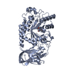 6or4C 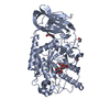 6orfC  6orgC  3eypS S: Starting model for refinement C: citing same article ( |
|---|---|
| Similar structure data |


-Links
 PDBj
PDBj

- Assembly
| Deposited unit | 
| ||||||||
|---|---|---|---|---|---|---|---|---|---|
| 1 |
| ||||||||
| Unit cell |
|
-Components
| #1: Protein | Mass: 50961.961 Da / Num. of mol.: 2 / Mutation: D171N, E215Q Source method: isolated from a genetically manipulated source Source: (gene. exp.) Streptococcus pneumoniae serotype 4 (strain ATCC BAA-334 / TIGR4) (bacteria)Strain: ATCC BAA-334 / TIGR4 / Gene: SP_2146 / Plasmid: pET28a / Production host: #2: Polysaccharide | 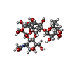  Source method: isolated from a genetically manipulated source Details: oligosaccharide with branches / References: Lewis Y antigen, alpha anomer #3: Chemical | ChemComp-EDO /   Mass: 62.068 Da / Num. of mol.: 14 Mass: 62.068 Da / Num. of mol.: 14Source method: isolated from a genetically manipulated source Formula: C2H6O2 #4: Water | ChemComp-HOH / |  Mass: 18.015 Da / Num. of mol.: 1031 / Source method: isolated from a natural source / Formula: H2O Mass: 18.015 Da / Num. of mol.: 1031 / Source method: isolated from a natural source / Formula: H2O |
|---|
-Experimental details
-Experiment
| Experiment | Method: X-RAY DIFFRACTION / Number of used crystals: 1 |
|---|
- Sample preparation
| Crystal | Density Matthews: 2.66 Å3/Da / Density % sol: 53.75 % |
|---|---|
| Crystal grow | Temperature: 291 K / Method: vapor diffusion, hanging drop / pH: 8.5 Details: 21 % PEG 4000, 0.22 M NaOAc, 1 mM DTT and 0.1 M Tris. |
-Data collection
| Diffraction | Mean temperature: 100 K / Serial crystal experiment: N |
|---|---|
| Diffraction source | Source: SYNCHROTRON / Site: SSRL  / Beamline: BL11-1 / Wavelength: 0.97945 Å / Beamline: BL11-1 / Wavelength: 0.97945 Å |
| Detector | Type: DECTRIS PILATUS 6M / Detector: PIXEL / Date: Jul 2, 2014 |
| Radiation | Protocol: SINGLE WAVELENGTH / Monochromatic (M) / Laue (L): M / Scattering type: x-ray |
| Radiation wavelength | Wavelength: 0.97945 Å / Relative weight: 1 |
| Reflection | Resolution: 1.62→27.84 Å / Num. obs: 131486 / % possible obs: 97.2 % / Redundancy: 4.5 % / CC1/2: 0.998 / Rmerge(I) obs: 0.05 / Rpim(I) all: 0.026 / Net I/σ(I): 17 |
| Reflection shell | Resolution: 1.62→1.71 Å / Redundancy: 4.5 % / Rmerge(I) obs: 0.337 / Mean I/σ(I) obs: 4.7 / Num. unique obs: 19408 / CC1/2: 0.882 / Rpim(I) all: 0.183 / % possible all: 98.4 |
-Phasing
| Phasing | Method: molecular replacement |
|---|
- Processing
| Software |
| |||||||||||||||||||||||||||||||||||||||||||||
|---|---|---|---|---|---|---|---|---|---|---|---|---|---|---|---|---|---|---|---|---|---|---|---|---|---|---|---|---|---|---|---|---|---|---|---|---|---|---|---|---|---|---|---|---|---|---|
| Refinement | Method to determine structure: MOLECULAR REPLACEMENT Starting model: 3EYP Resolution: 1.62→27.84 Å / Cor.coef. Fo:Fc: 0.969 / Cor.coef. Fo:Fc free: 0.954 / SU B: 1.784 / SU ML: 0.061 / Cross valid method: THROUGHOUT / σ(F): 0 / ESU R: 0.087 / ESU R Free: 0.089 / Stereochemistry target values: MAXIMUM LIKELIHOOD / Details: U VALUES : REFINED INDIVIDUALLY
| |||||||||||||||||||||||||||||||||||||||||||||
| Solvent computation | Ion probe radii: 0.8 Å / Shrinkage radii: 0.8 Å / VDW probe radii: 1.2 Å / Solvent model: MASK | |||||||||||||||||||||||||||||||||||||||||||||
| Displacement parameters | Biso max: 64.14 Å2 / Biso mean: 21.902 Å2 / Biso min: 8.41 Å2
| |||||||||||||||||||||||||||||||||||||||||||||
| Refinement step | Cycle: final / Resolution: 1.62→27.84 Å
| |||||||||||||||||||||||||||||||||||||||||||||
| Refine LS restraints |
| |||||||||||||||||||||||||||||||||||||||||||||
| LS refinement shell | Resolution: 1.619→1.661 Å / Rfactor Rfree error: 0 / Total num. of bins used: 20
|
