 Movie
Movie Controller

[English] 日本語
 Yorodumi
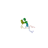
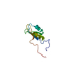
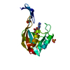
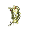
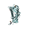
- PDB-6oqj: SOLUTION STRUCTURE OF THE COMPLEX OF MUTANT VEK50[RH1/AA] AND PLA... -
+ Open data

- Basic information
| Entry | Database: PDB / ID: 6oqj | ||||||
|---|---|---|---|---|---|---|---|
| Title | SOLUTION STRUCTURE OF THE COMPLEX OF MUTANT VEK50[RH1/AA] AND PLASMINOGEN KRINGLE 2 | ||||||
 Components Components |
| ||||||
 Keywords Keywords | PROTEIN BINDING / PLASMINOGEN BINDING PROTEIN / BLOOD CLOTTING | ||||||
| Function / homology |  Function and homology information Function and homology informationplasmin / trans-synaptic signaling by BDNF, modulating synaptic transmission / trophoblast giant cell differentiation / tissue remodeling / tissue regeneration / Signaling by PDGF / positive regulation of fibrinolysis / mononuclear cell migration / negative regulation of cell-cell adhesion mediated by cadherin / protein antigen binding ...plasmin / trans-synaptic signaling by BDNF, modulating synaptic transmission / trophoblast giant cell differentiation / tissue remodeling / tissue regeneration / Signaling by PDGF / positive regulation of fibrinolysis / mononuclear cell migration / negative regulation of cell-cell adhesion mediated by cadherin / protein antigen binding / myoblast differentiation / Dissolution of Fibrin Clot / labyrinthine layer blood vessel development / muscle cell cellular homeostasis / plasminogen activation / biological process involved in interaction with symbiont / Activation of Matrix Metalloproteinases / extracellular matrix disassembly / negative regulation of fibrinolysis / negative regulation of cell-substrate adhesion / apolipoprotein binding / positive regulation of blood vessel endothelial cell migration / fibrinolysis / Degradation of the extracellular matrix / platelet alpha granule lumen / serine-type peptidase activity / protein processing / Schaffer collateral - CA1 synapse / Regulation of Insulin-like Growth Factor (IGF) transport and uptake by Insulin-like Growth Factor Binding Proteins (IGFBPs) / kinase binding / blood coagulation / Platelet degranulation / protein-folding chaperone binding / extracellular matrix / protease binding / endopeptidase activity / blood microparticle / negative regulation of cell population proliferation / serine-type endopeptidase activity / external side of plasma membrane / protein domain specific binding / signaling receptor binding / glutamatergic synapse / enzyme binding / cell surface / proteolysis / : / extracellular exosome / extracellular region / plasma membrane Similarity search - Function | ||||||
| Biological species |  Homo sapiens (human) Homo sapiens (human) Streptococcus pyogenes (bacteria) Streptococcus pyogenes (bacteria) | ||||||
| Method | SOLUTION NMR / simulated annealing | ||||||
 Authors Authors | Yuan, Y. / Castellino, F.J. | ||||||
| Funding support |  United States, 1items United States, 1items
| ||||||
 Citation Citation | Journal: J.Struct.Biol. / Year: 2019 Title: Solution structural model of the complex of the binding regions of human plasminogen with its M-protein receptor from Streptococcus pyogenes. Authors: Yuan, Y. / Ayinuola, Y.A. / Singh, D. / Ayinuola, O. / Mayfield, J.A. / Quek, A. / Whisstock, J.C. / Law, R.H.P. / Lee, S.W. / Ploplis, V.A. / Castellino, F.J. | ||||||
| History |
|
- Structure visualization
| Structure viewer | Molecule: MolmilJmol/JSmol |
|---|
- Downloads & links
-Download
| PDBx/mmCIF format | 6oqj.cif.gz | 434 KB | Display | PDBx/mmCIF format |
|---|---|---|---|---|
| PDB format | pdb6oqj.ent.gz | 359.9 KB | Display | PDB format |
| PDBx/mmJSON format | 6oqj.json.gz | Tree view | PDBx/mmJSON format | |
| Others |  Other downloads Other downloads |
-Validation report
| Arichive directory | https://data.pdbj.org/pub/pdb/validation_reports/oq/6oqjftp://data.pdbj.org/pub/pdb/validation_reports/oq/6oqj | HTTPS FTP |
|---|
-Related structure data
| Related structure data |  6okwC 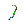 6okxC 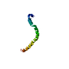 6okyC 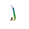 6oq9C 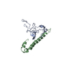 6oqkC C: citing same article ( |
|---|---|
| Similar structure data | |
| Other databases |
|


-Links
 PDBj
PDBj


- Assembly
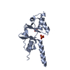
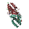
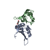
| Deposited unit | 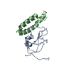
| |||||||||
|---|---|---|---|---|---|---|---|---|---|---|
| 1 |
| |||||||||
| NMR ensembles |
|
-Components
| #1: Protein | Mass: 10166.340 Da / Num. of mol.: 1 Source method: isolated from a genetically manipulated source Source: (gene. exp.) Homo sapiens (human) / Plasmid: PPIC9K / Production host:  Komagataella pastoris (fungus) / References: UniProt: P00747*PLUS Komagataella pastoris (fungus) / References: UniProt: P00747*PLUS |
|---|---|
| #2: Protein | Mass: 6020.606 Da / Num. of mol.: 1 / Fragment: residues 85-134 / Mutation: R19A, H20A Source method: isolated from a genetically manipulated source Details: AP53 / Source: (gene. exp.) Streptococcus pyogenes (bacteria) / Gene: pam, emm / Plasmid: pET15b / Production host: |
-Experimental details
-Experiment
| Experiment | Method: SOLUTION NMR | ||||||||||||||||||||||||||||||||||||||||||||||||||||||
|---|---|---|---|---|---|---|---|---|---|---|---|---|---|---|---|---|---|---|---|---|---|---|---|---|---|---|---|---|---|---|---|---|---|---|---|---|---|---|---|---|---|---|---|---|---|---|---|---|---|---|---|---|---|---|---|
| NMR experiment |
|
 HSQC
HSQC- Sample preparation
| Details |
| ||||||||||||||||||||||||||||||||||||||||||||
|---|---|---|---|---|---|---|---|---|---|---|---|---|---|---|---|---|---|---|---|---|---|---|---|---|---|---|---|---|---|---|---|---|---|---|---|---|---|---|---|---|---|---|---|---|---|
| Sample |
| ||||||||||||||||||||||||||||||||||||||||||||
| Sample conditions | Ionic strength: 20 mM / Label: conditions_1 / pH: 6.8 / Pressure: 1 atm / Temperature: 298 K |
-NMR measurement
| NMR spectrometer | Type: Bruker AVANCE / Manufacturer: Bruker / Model: AVANCE / Field strength: 800 MHz |
|---|
- Processing
| NMR software |
| |||||||||||||||||||||
|---|---|---|---|---|---|---|---|---|---|---|---|---|---|---|---|---|---|---|---|---|---|---|
| Refinement |
| |||||||||||||||||||||
| NMR representative | Selection criteria: lowest energy | |||||||||||||||||||||
| NMR ensemble | Conformer selection criteria: structures with the lowest energy Conformers calculated total number: 200 / Conformers submitted total number: 10 |
