 Movie
Movie Controller

[English] 日本語
 Yorodumi
- PDB-3x0n: ADP ribose pyrophosphatase from Thermus thermophilus HB8 in ESM-s... -
+ Open data

- Basic information
| Entry | Database: PDB / ID: 3x0n | ||||||
|---|---|---|---|---|---|---|---|
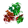
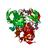
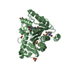
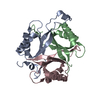
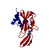
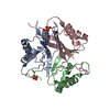
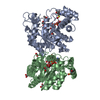
| Title | ADP ribose pyrophosphatase from Thermus thermophilus HB8 in ESM-state at reaction time of 6 min | ||||||
 Components Components | ADP-ribose pyrophosphatase | ||||||
 Keywords Keywords | HYDROLASE / Nudix motif / ADP ribose hydrolase / ADP ribose / Cytosol | ||||||
| Function / homology |  Function and homology information Function and homology informationpyrophosphatase activity / nucleoside phosphate metabolic process / ribose phosphate metabolic process / metal ion binding / cytosol Similarity search - Function | ||||||
| Biological species |   Thermus thermophilus HB8 (bacteria) Thermus thermophilus HB8 (bacteria) | ||||||
| Method |  X-RAY DIFFRACTION / SYNCHROTRON / MOLECULAR REPLACEMENT / Resolution: 1.12 Å X-RAY DIFFRACTION / SYNCHROTRON / MOLECULAR REPLACEMENT / Resolution: 1.12 Å | ||||||
 Authors Authors | Furuike, Y. / Akita, Y. / Miyahara, I. / Kamiya, N. | ||||||
 Citation Citation | Journal: Biochemistry / Year: 2016 Title: ADP-Ribose Pyrophosphatase Reaction in Crystalline State Conducted by Consecutive Binding of Two Manganese(II) Ions as Cofactors Authors: Furuike, Y. / Akita, Y. / Miyahara, I. / Kamiya, N. | ||||||
| History |
|
- Structure visualization
| Structure viewer | Molecule: MolmilJmol/JSmol |
|---|
- Downloads & links
-Download
| PDBx/mmCIF format | 3x0n.cif.gz | 89 KB | Display | PDBx/mmCIF format |
|---|---|---|---|---|
| PDB format | pdb3x0n.ent.gz | 65.8 KB | Display | PDB format |
| PDBx/mmJSON format | 3x0n.json.gz | Tree view | PDBx/mmJSON format | |
| Others |  Other downloads Other downloads |
-Validation report
| Arichive directory | https://data.pdbj.org/pub/pdb/validation_reports/x0/3x0nftp://data.pdbj.org/pub/pdb/validation_reports/x0/3x0n | HTTPS FTP |
|---|
-Related structure data
| Related structure data | 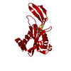 3x0iSC 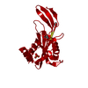 3x0jC 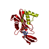 3x0kC 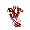 3x0lC 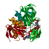 3x0mC  3x0oC  3x0pC 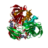 3x0qC  3x0rC 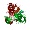 3x0sC S: Starting model for refinement C: citing same article ( |
|---|---|
| Similar structure data |


-Links
 PDBj
PDBj


- Assembly
| Deposited unit | 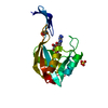
| ||||||||
|---|---|---|---|---|---|---|---|---|---|
| 1 | 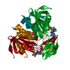
| ||||||||
| Unit cell |
| ||||||||
| Components on special symmetry positions |
|
-Components
-Protein , 1 types, 1 molecules A
| #1: Protein | Mass: 19287.912 Da / Num. of mol.: 1 Source method: isolated from a genetically manipulated source Source: (gene. exp.) Thermus thermophilus HB8 (bacteria) / Strain: HB8 / Gene: TTHA0528 / Production host: |
|---|
-Non-polymers , 5 types, 127 molecules 


| #2: Chemical | ChemComp-AR6 / [( Mass: 559.316 Da / Num. of mol.: 1 / Source method: obtained synthetically / Formula: C15H23N5O14P2 Mass: 559.316 Da / Num. of mol.: 1 / Source method: obtained synthetically / Formula: C15H23N5O14P2 |
|---|---|
| #3: Chemical | ChemComp-MN /  Mass: 54.938 Da / Num. of mol.: 1 / Source method: obtained synthetically / Formula: Mn Mass: 54.938 Da / Num. of mol.: 1 / Source method: obtained synthetically / Formula: Mn |
| #4: Chemical | ChemComp-GOL /  Mass: 92.094 Da / Num. of mol.: 1 / Source method: obtained synthetically / Formula: C3H8O3 Mass: 92.094 Da / Num. of mol.: 1 / Source method: obtained synthetically / Formula: C3H8O3 |
| #5: Chemical | ChemComp-SO4 /  Mass: 96.063 Da / Num. of mol.: 1 / Source method: obtained synthetically / Formula: SO4 Mass: 96.063 Da / Num. of mol.: 1 / Source method: obtained synthetically / Formula: SO4 |
| #6: Water | ChemComp-HOH / Mass: 18.015 Da / Num. of mol.: 123 / Source method: isolated from a natural source / Formula: H2O |
-Experimental details
-Experiment
| Experiment | Method: X-RAY DIFFRACTION / Number of used crystals: 1 |
|---|
- Sample preparation
| Crystal | Density Matthews: 2.2 Å3/Da / Density % sol: 44.04 % |
|---|---|
| Crystal grow | Temperature: 298 K / Method: vapor diffusion, hanging drop / pH: 4.6 Details: 0.24M Acetate-Sodium acetate buffer, 0.32M Ammonium sulfate, 30%(w/v) Glycerol, 8%(w/v) PEG 20000, pH 4.6, VAPOR DIFFUSION, HANGING DROP, temperature 298K |
-Data collection
| Diffraction | Mean temperature: 100 K | |||||||||||||||||||||||||||||||||
|---|---|---|---|---|---|---|---|---|---|---|---|---|---|---|---|---|---|---|---|---|---|---|---|---|---|---|---|---|---|---|---|---|---|---|
| Diffraction source | Source: SYNCHROTRON / Site: SPring-8  / Beamline: BL38B1 / Wavelength: 0.9 Å / Beamline: BL38B1 / Wavelength: 0.9 Å | |||||||||||||||||||||||||||||||||
| Detector | Type: ADSC QUANTUM 315 / Detector: CCD / Date: Oct 10, 2012 | |||||||||||||||||||||||||||||||||
| Radiation | Monochromator: Fixed exit Si (111) double crystal monochromator Protocol: SINGLE WAVELENGTH / Monochromatic (M) / Laue (L): M / Scattering type: x-ray | |||||||||||||||||||||||||||||||||
| Radiation wavelength | Wavelength: 0.9 Å / Relative weight: 1 | |||||||||||||||||||||||||||||||||
| Reflection | Resolution: 1.12→30 Å / Num. all: 66244 / Num. obs: 66244 / % possible obs: 99.8 % / Observed criterion σ(F): 0 / Observed criterion σ(I): 0 | |||||||||||||||||||||||||||||||||
| Reflection shell |
|
- Processing
| Software |
| ||||||||||||
|---|---|---|---|---|---|---|---|---|---|---|---|---|---|
| Refinement | Method to determine structure: MOLECULAR REPLACEMENT Starting model: 3X0I Resolution: 1.12→30 Å / σ(F): 0 / Stereochemistry target values: Engh & Huber /
| ||||||||||||
| Refinement step | Cycle: LAST / Resolution: 1.12→30 Å
| ||||||||||||
| Refine LS restraints |
|
