 Movie
Movie Controller

[English] 日本語
 Yorodumi
- PDB-6o6z: Crystal structure of Csm6 H381A in complex with cA4 by cocrystall... -
+ Open data

- Basic information
| Entry | Database: PDB / ID: 6o6z | ||||||
|---|---|---|---|---|---|---|---|
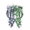
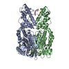
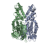
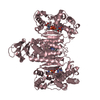
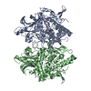
| Title | Crystal structure of Csm6 H381A in complex with cA4 by cocrystallization of cA4 and Csm6 | ||||||
 Components Components | Csm6 | ||||||
 Keywords Keywords | IMMUNE SYSTEM / Type III-A CRISPR-Cas system / Csm6 | ||||||
| Function / homology | CRISPR system endoribonuclease Csx1, HEPN domain / CRISPR system endoribonuclease Csx1 / CRISPR-associated protein DxTHG, conserved site / : / CRISPR system endoribonuclease Csx1, CARF domain / : / Chem-LQJ / CRISPR system endoribonuclease Csx1 CARF domain-containing protein Function and homology information Function and homology information | ||||||
| Biological species |   Thermococcus onnurineus (archaea) Thermococcus onnurineus (archaea) | ||||||
| Method |  X-RAY DIFFRACTION / SYNCHROTRON / MOLECULAR REPLACEMENT / Resolution: 2.1 Å X-RAY DIFFRACTION / SYNCHROTRON / MOLECULAR REPLACEMENT / Resolution: 2.1 Å | ||||||
 Authors Authors | Jia, N. / Patel, D.J. | ||||||
 Citation Citation | Journal: Mol.Cell / Year: 2019 Title: CRISPR-Cas III-A Csm6 CARF Domain Is a Ring Nuclease Triggering Stepwise cA4Cleavage with ApA>p Formation Terminating RNase Activity. Authors: Jia, N. / Jones, R. / Yang, G. / Ouerfelli, O. / Patel, D.J. | ||||||
| History |
|
- Structure visualization
| Structure viewer | Molecule: MolmilJmol/JSmol |
|---|
- Downloads & links
-Download
| PDBx/mmCIF format | 6o6z.cif.gz | 185.8 KB | Display | PDBx/mmCIF format |
|---|---|---|---|---|
| PDB format | pdb6o6z.ent.gz | 147.2 KB | Display | PDB format |
| PDBx/mmJSON format | 6o6z.json.gz | Tree view | PDBx/mmJSON format | |
| Others |  Other downloads Other downloads |
-Validation report
| Arichive directory | https://data.pdbj.org/pub/pdb/validation_reports/o6/6o6zftp://data.pdbj.org/pub/pdb/validation_reports/o6/6o6z | HTTPS FTP |
|---|
-Related structure data
| Related structure data |  6o6sC  6o6tC 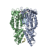 6o6vC  6o6xC 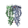 6o6yC  6o70C 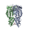 6o71C 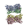 6ov0C C: citing same article ( |
|---|---|
| Similar structure data |


-Links
 PDBj
PDBj- Assembly
| Deposited unit | 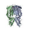
| ||||||||
|---|---|---|---|---|---|---|---|---|---|
| 1 |
| ||||||||
| Unit cell |
|
-Components
| #1: Protein | Mass: 49728.047 Da / Num. of mol.: 2 / Mutation: H381A Source method: isolated from a genetically manipulated source Source: (gene. exp.) Thermococcus onnurineus (strain NA1) (archaea)Strain: NA1 / Gene: TON_0898 / Production host:  #2: Chemical |   Mass: 658.412 Da / Num. of mol.: 2 / Source method: obtained synthetically / Formula: C20H24N10O12P2 Mass: 658.412 Da / Num. of mol.: 2 / Source method: obtained synthetically / Formula: C20H24N10O12P2#3: Water | ChemComp-HOH / |  Mass: 18.015 Da / Num. of mol.: 150 / Source method: isolated from a natural source / Formula: H2O Mass: 18.015 Da / Num. of mol.: 150 / Source method: isolated from a natural source / Formula: H2ONonpolymer details | LQJ is a result of cA4 cleavage occurred during crystallization | |
|---|
-Experimental details
-Experiment
| Experiment | Method: X-RAY DIFFRACTION / Number of used crystals: 1 |
|---|
- Sample preparation
| Crystal | Density Matthews: 2.52 Å3/Da / Density % sol: 51.23 % |
|---|---|
| Crystal grow | Temperature: 293 K / Method: vapor diffusion, hanging drop / pH: 6.2 Details: 10% PEG 8K, 0.2 M NaCl, 0.1 M Na/K phosphate pH 6.2 |
-Data collection
| Diffraction | Mean temperature: 100 K / Serial crystal experiment: N |
|---|---|
| Diffraction source | Source: SYNCHROTRON / Site: APS  / Beamline: 24-ID-E / Wavelength: 0.9791 Å / Beamline: 24-ID-E / Wavelength: 0.9791 Å |
| Detector | Type: DECTRIS PILATUS 6M / Detector: PIXEL / Date: Dec 11, 2018 |
| Radiation | Protocol: SINGLE WAVELENGTH / Monochromatic (M) / Laue (L): M / Scattering type: x-ray |
| Radiation wavelength | Wavelength: 0.9791 Å / Relative weight: 1 |
| Reflection | Resolution: 2.1→50 Å / Num. obs: 59191 / % possible obs: 99.4 % / Redundancy: 10.9 % / Rpim(I) all: 0.055 / Net I/σ(I): 16.9 |
| Reflection shell | Resolution: 2.1→2.18 Å / Num. unique obs: 5719 |
- Processing
| Software |
| ||||||||||||||||||||||||||||||||||||||||||||||||||||||||||||||||||||||||||||||||||||||||||||||||||||||||||||||||||||||||||||||||||||||||||||||||||||||||||||||||||||||||||||||||||||||
|---|---|---|---|---|---|---|---|---|---|---|---|---|---|---|---|---|---|---|---|---|---|---|---|---|---|---|---|---|---|---|---|---|---|---|---|---|---|---|---|---|---|---|---|---|---|---|---|---|---|---|---|---|---|---|---|---|---|---|---|---|---|---|---|---|---|---|---|---|---|---|---|---|---|---|---|---|---|---|---|---|---|---|---|---|---|---|---|---|---|---|---|---|---|---|---|---|---|---|---|---|---|---|---|---|---|---|---|---|---|---|---|---|---|---|---|---|---|---|---|---|---|---|---|---|---|---|---|---|---|---|---|---|---|---|---|---|---|---|---|---|---|---|---|---|---|---|---|---|---|---|---|---|---|---|---|---|---|---|---|---|---|---|---|---|---|---|---|---|---|---|---|---|---|---|---|---|---|---|---|---|---|---|---|
| Refinement | Method to determine structure: MOLECULAR REPLACEMENT Starting model: 6O6Z Resolution: 2.1→48.35 Å / Cor.coef. Fo:Fc: 0.958 / Cor.coef. Fo:Fc free: 0.947 / SU B: 5.255 / SU ML: 0.133 / Cross valid method: THROUGHOUT / ESU R: 0.209 / ESU R Free: 0.169 / Details: HYDROGENS HAVE BEEN ADDED IN THE RIDING POSITIONS
| ||||||||||||||||||||||||||||||||||||||||||||||||||||||||||||||||||||||||||||||||||||||||||||||||||||||||||||||||||||||||||||||||||||||||||||||||||||||||||||||||||||||||||||||||||||||
| Solvent computation | Ion probe radii: 0.8 Å / Shrinkage radii: 0.8 Å / VDW probe radii: 1.2 Å | ||||||||||||||||||||||||||||||||||||||||||||||||||||||||||||||||||||||||||||||||||||||||||||||||||||||||||||||||||||||||||||||||||||||||||||||||||||||||||||||||||||||||||||||||||||||
| Displacement parameters | Biso mean: 40.976 Å2
| ||||||||||||||||||||||||||||||||||||||||||||||||||||||||||||||||||||||||||||||||||||||||||||||||||||||||||||||||||||||||||||||||||||||||||||||||||||||||||||||||||||||||||||||||||||||
| Refinement step | Cycle: 1 / Resolution: 2.1→48.35 Å
| ||||||||||||||||||||||||||||||||||||||||||||||||||||||||||||||||||||||||||||||||||||||||||||||||||||||||||||||||||||||||||||||||||||||||||||||||||||||||||||||||||||||||||||||||||||||
| Refine LS restraints |
|