[English] 日本語
Yorodumi
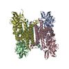
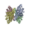
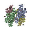
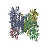
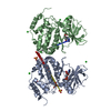
- PDB-6o6q: Crystal structure of Cka1p, a casein kinase 2 alpha ortholog from... -

+
Open data


ID or keywords:

Loading...

-
Basic information

Entry
Database: PDB / ID: 6o6q
TitleCrystal structure of Cka1p, a casein kinase 2 alpha ortholog from Candida albicans
ComponentsCasein kinase 2 catalytic subunit
KeywordsTRANSFERASE / kinase / alpha/beta fold / fungus / pathogenesis / National Institute of Allergy and Infectious Diseases / NIAID / National Institutes of Health / NIH / structural genomics / Center for Structural Genomics of Infectious Diseases / CSGID
Function / homology
Function and homology information


regulation of transcription by RNA polymerase I / regulation of transcription by RNA polymerase III / protein kinase CK2 complex / peptidyl-threonine phosphorylation / peptidyl-serine phosphorylation / regulation of cell cycle / protein serine/threonine kinase activity / DNA damage response / ATP binding / nucleus / cytosol
Similarity search - Function
Transferase(Phosphotransferase) domain 1 / Transferase(Phosphotransferase); domain 1 / Phosphorylase Kinase; domain 1 / Phosphorylase Kinase; domain 1 / Serine/threonine-protein kinase, active site / Serine/Threonine protein kinases active-site signature. / Protein kinase domain / Serine/Threonine protein kinases, catalytic domain / Protein kinase, ATP binding site / Protein kinases ATP-binding region signature. ...Transferase(Phosphotransferase) domain 1 / Transferase(Phosphotransferase); domain 1 / Phosphorylase Kinase; domain 1 / Phosphorylase Kinase; domain 1 / Serine/threonine-protein kinase, active site / Serine/Threonine protein kinases active-site signature. / Protein kinase domain / Serine/Threonine protein kinases, catalytic domain / Protein kinase, ATP binding site / Protein kinases ATP-binding region signature. / Protein kinase domain profile. / Protein kinase domain / Protein kinase-like domain superfamily / 2-Layer Sandwich / Orthogonal Bundle / Mainly Alpha / Alpha Beta
Similarity search - Domain/homology
ADENOSINE MONOPHOSPHATE / Casein kinase 2 catalytic subunit
Similarity search - Component
Biological speciesCandida albicans (yeast)
MethodX-RAY DIFFRACTION / MOLECULAR REPLACEMENT / Resolution: 2.7 Å
AuthorsStogios, P.J. / Evdokimova, E. / Di Leo, R. / Savchenko, A. / Center for Structural Genomics of Infectious Diseases (CSGID)
Funding support United States, 1items
OrganizationGrant numberCountry
National Institutes of Health/National Institute Of Allergy and Infectious Diseases (NIH/NIAID)HHSN272201700060C United States
CitationJournal: To Be Published
Title: Crystal structure of Cka1p, a casein kinase 2 alpha ortholog from Candida albicans
Authors: Stogios, P.J.
History
DepositionMar 7, 2019Deposition site: RCSB / Processing site: RCSB
Revision 1.0Apr 3, 2019Provider: repository / Type: Initial release
Revision 1.1Dec 18, 2019Group: Author supporting evidence / Category: pdbx_audit_support / Item: _pdbx_audit_support.funding_organization
Revision 1.2Oct 11, 2023Group: Data collection / Database references / Refinement description
Category: chem_comp_atom / chem_comp_bond ...chem_comp_atom / chem_comp_bond / database_2 / pdbx_initial_refinement_model
Item: _database_2.pdbx_DOI / _database_2.pdbx_database_accession

-
Structure visualization

Structure viewerMolecule:
MolmilJmol/JSmol

Downloads & links

-
Assembly

Deposited unit
A: Casein kinase 2 catalytic subunit
B: Casein kinase 2 catalytic subunit
hetero molecules


Theoretical massNumber of molelcules
Total (without water)81,49317
Polymers80,0462
Non-polymers1,44615
Water3,153175
1
A: Casein kinase 2 catalytic subunit
B: Casein kinase 2 catalytic subunit
hetero molecules

A: Casein kinase 2 catalytic subunit
B: Casein kinase 2 catalytic subunit
hetero molecules


Theoretical massNumber of molelcules
Total (without water)162,98534
Polymers160,0924
Non-polymers2,89330
Water724
TypeNameSymmetry operationNumber
identity operation1_555x,y,z1
crystal symmetry operation8_555x-y,-y,-z1
Buried area12520 Å2
ΔGint-209 kcal/mol
Surface area58710 Å2
MethodPISA
Unit cell
Length a, b, c (Å)125.690, 125.690, 229.719
Angle α, β, γ (deg.)90.00, 90.00, 120.00
Int Tables number178
Space group name H-MP6122
Components on special symmetry positions
IDModelComponents
11A-573-

HOH

-
Components

-
Protein , 1 types, 2 molecules AB

#1: Protein Casein kinase 2 catalytic subunit


Mass: 40023.035 Da / Num. of mol.: 2
Source method: isolated from a genetically manipulated source
Source: (gene. exp.) Candida albicans (strain SC5314 / ATCC MYA-2876) (yeast)
Strain: SC5314 / ATCC MYA-2876 / Gene: CKA1, CAALFM_CR10660WA, orf19.7652 / Plasmid: pMCSG68SBPTEV / Production host: Escherichia coli BL21(DE3) (bacteria) / Strain (production host): BL21(DE3)-Gold / References: UniProt: A0A1D8PUA2

-
Non-polymers , 5 types, 190 molecules

#2: Chemical ChemComp-AMP / ADENOSINE MONOPHOSPHATE


Mass: 347.221 Da / Num. of mol.: 2 / Source method: obtained synthetically / Formula: C10H14N5O7P / Comment: AMP*YM
#3: Chemical ChemComp-SO4 / SULFATE ION


Mass: 96.063 Da / Num. of mol.: 2 / Source method: obtained synthetically / Formula: SO4
#4: Chemical
ChemComp-CL / CHLORIDE ION


Mass: 35.453 Da / Num. of mol.: 8 / Source method: obtained synthetically / Formula: Cl
#5: Chemical ChemComp-GOL / GLYCEROL / GLYCERIN / PROPANE-1,2,3-TRIOL


Mass: 92.094 Da / Num. of mol.: 3 / Source method: obtained synthetically / Formula: C3H8O3
#6: Water ChemComp-HOH / water


Mass: 18.015 Da / Num. of mol.: 175 / Source method: isolated from a natural source / Formula: H2O

-
Experimental details

-
Experiment

ExperimentMethod: X-RAY DIFFRACTION / Number of used crystals: 1

-
Sample preparation

CrystalDensity Matthews: 3.27 Å3/Da / Density % sol: 62.41 %
Crystal growTemperature: 298 K / Method: vapor diffusion, sitting drop / pH: 8
Details: 2 M ammonium sulfate, 0.1 M sodium citrate pH 6.1, 1 mM AMP-PNP

-
Data collection

DiffractionMean temperature: 100 K / Serial crystal experiment: N
Diffraction sourceSource: ROTATING ANODE / Type: RIGAKU MICROMAX-007 HF / Wavelength: 1.5418 Å
DetectorType: RIGAKU RAXIS IV / Detector: IMAGE PLATE / Date: Aug 30, 2018
RadiationProtocol: SINGLE WAVELENGTH / Monochromatic (M) / Laue (L): M / Scattering type: x-ray
Radiation wavelengthWavelength: 1.5418 Å / Relative weight: 1
ReflectionResolution: 2.66→35 Å / Num. obs: 31301 / % possible obs: 98.9 % / Redundancy: 18.3 % / Rmerge(I) obs: 0.184 / Rpim(I) all: 0.043 / Net I/σ(I): 18.57
Reflection shellResolution: 2.66→2.71 Å / Redundancy: 9.3 % / Rmerge(I) obs: 1.004 / Mean I/σ(I) obs: 1.23 / Num. unique obs: 1254 / CC1/2: 0.787 / Rpim(I) all: 0.305 / % possible all: 82.1

-
Processing

Software
NameVersionClassification
PHENIX1.14_3260refinement
HKL-3000data reduction
HKL-3000data scaling
PHENIXphasing
PHENIXmodel building
Cootmodel building
RefinementMethod to determine structure: MOLECULAR REPLACEMENT
Starting model: 3bqc
Resolution: 2.7→34.598 Å / SU ML: 0.33 / Cross valid method: FREE R-VALUE / σ(F): 1.33 / Phase error: 30.12
RfactorNum. reflection% reflectionSelection details
Rfree0.2873 2360 5.02 %RANDOM
Rwork0.2335 ---
obs0.2361 47037 84.39 %-
Solvent computationShrinkage radii: 0.9 Å / VDW probe radii: 1.11 Å
Refinement stepCycle: LAST / Resolution: 2.7→34.598 Å
ProteinNucleic acidLigandSolventTotal
Num. atoms5498 0 82 175 5755
Refine LS restraints
Refine-IDTypeDev idealNumber
X-RAY DIFFRACTIONf_bond_d0.0045706
X-RAY DIFFRACTIONf_angle_d0.6477696
X-RAY DIFFRACTIONf_dihedral_angle_d21.3252169
X-RAY DIFFRACTIONf_chiral_restr0.054805
X-RAY DIFFRACTIONf_plane_restr0.003962
LS refinement shell
Resolution (Å)Rfactor RfreeNum. reflection RfreeRfactor RworkNum. reflection RworkRefine-ID% reflection obs (%)
2.7001-2.75520.4108640.28321076X-RAY DIFFRACTION35
2.7552-2.81510.3123680.30471341X-RAY DIFFRACTION43
2.8151-2.88050.3188960.30981838X-RAY DIFFRACTION59
2.8805-2.95250.34221150.3152185X-RAY DIFFRACTION70
2.9525-3.03230.40131230.32952348X-RAY DIFFRACTION76
3.0323-3.12150.33811350.29552515X-RAY DIFFRACTION81
3.1215-3.22210.32291450.29852709X-RAY DIFFRACTION87
3.2221-3.33720.35691590.27172985X-RAY DIFFRACTION96
3.3372-3.47070.31791650.25333081X-RAY DIFFRACTION99
3.4707-3.62850.29591620.23183112X-RAY DIFFRACTION99
3.6285-3.81960.28031570.21743107X-RAY DIFFRACTION99
3.8196-4.05860.2811610.2043045X-RAY DIFFRACTION98
4.0586-4.37140.23571550.18063061X-RAY DIFFRACTION98
4.3714-4.81020.2241610.17883060X-RAY DIFFRACTION98
4.8102-5.50390.24811620.20183069X-RAY DIFFRACTION99
5.5039-6.92520.32021660.25323078X-RAY DIFFRACTION99
6.9252-34.60120.25251660.23253067X-RAY DIFFRACTION98
Refinement TLS params.

Method: refined / Refine-ID: X-RAY DIFFRACTION

IDL112)L122)L132)L222)L232)L332)S11 (Å °)S12 (Å °)S13 (Å °)S21 (Å °)S22 (Å °)S23 (Å °)S31 (Å °)S32 (Å °)S33 (Å °)T112)T122)T132)T222)T232)T332)Origin x (Å)Origin y (Å)Origin z (Å)
11.82230.18190.95661.67810.25893.7075-0.0739-0.10710.1079-0.06180.07380.1489-0.0138-0.335-0.00690.2091-0.0103-0.00080.18460.0460.25942.607629.5052-6.9179
20.55740.1333-0.12890.9896-0.04781.3591-0.0649-0.0226-0.29920.46610.3324-0.16080.93061.09410.01130.90510.2598-0.10690.512-0.15370.588175.2078-4.449423.1793
31.94860.06740.69432.26530.1233.8914-0.00150.33960.06770.14540.1264-0.1980.14440.880.02590.24810.0635-0.03590.3267-0.07920.263574.053616.946521.311
40.49460.1880.11042.1042-0.83151.64080.09010.0629-0.0542-0.64690.11270.44120.5878-0.40480.00060.7655-0.0191-0.05090.39210.04150.464542.40789.9343-17.7681
Refinement TLS group
IDRefine-IDRefine TLS-IDSelection details
1X-RAY DIFFRACTION1chain 'A' and (resid 134 through 333 )
2X-RAY DIFFRACTION2chain 'B' and (resid 5 through 91 )
3X-RAY DIFFRACTION3chain 'B' and (resid 92 through 333 )
4X-RAY DIFFRACTION4chain 'A' and (resid 4 through 133 )

+
About Yorodumi

-
News

-
Feb 9, 2022. New format data for meta-information of EMDB entries

New format data for meta-information of EMDB entries

  • Version 3 of the EMDB header file is now the official format.
  • The previous official version 1.9 will be removed from the archive.

Related info.:EMDB header

External links:wwPDB to switch to version 3 of the EMDB data model

-
Aug 12, 2020. Covid-19 info

Covid-19 info

URL: https://pdbjlvh1.pdbj.org/emnavi/covid19.php

New page: Covid-19 featured information page in EM Navigator.

Related info.:Covid-19 info / Mar 5, 2020. Novel coronavirus structure data

+
Mar 5, 2020. Novel coronavirus structure data

Novel coronavirus structure data

Related info.:Yorodumi Speices / Aug 12, 2020. Covid-19 info

External links:COVID-19 featured content - PDBj / Molecule of the Month (242):Coronavirus Proteases

+
Jan 31, 2019. EMDB accession codes are about to change! (news from PDBe EMDB page)

EMDB accession codes are about to change! (news from PDBe EMDB page)

  • The allocation of 4 digits for EMDB accession codes will soon come to an end. Whilst these codes will remain in use, new EMDB accession codes will include an additional digit and will expand incrementally as the available range of codes is exhausted. The current 4-digit format prefixed with “EMD-” (i.e. EMD-XXXX) will advance to a 5-digit format (i.e. EMD-XXXXX), and so on. It is currently estimated that the 4-digit codes will be depleted around Spring 2019, at which point the 5-digit format will come into force.
  • The EM Navigator/Yorodumi systems omit the EMD- prefix.

Related info.:Q: What is EMD? / ID/Accession-code notation in Yorodumi/EM Navigator

External links:EMDB Accession Codes are Changing Soon! / Contact to PDBj

+
Jul 12, 2017. Major update of PDB

Major update of PDB

  • wwPDB released updated PDB data conforming to the new PDBx/mmCIF dictionary.
  • This is a major update changing the version number from 4 to 5, and with Remediation, in which all the entries are updated.
  • In this update, many items about electron microscopy experimental information are reorganized (e.g. em_software).
  • Now, EM Navigator and Yorodumi are based on the updated data.

External links:wwPDB Remediation / Enriched Model Files Conforming to OneDep Data Standards Now Available in the PDB FTP Archive

-
Yorodumi

Thousand views of thousand structures

  • Yorodumi is a browser for structure data from EMDB, PDB, SASBDB, etc.
  • This page is also the successor to EM Navigator detail page, and also detail information page/front-end page for Omokage search.
  • The word "yorodu" (or yorozu) is an old Japanese word meaning "ten thousand". "mi" (miru) is to see.

Related info.:EMDB / PDB / SASBDB / Comparison of 3 databanks / Yorodumi Search / Aug 31, 2016. New EM Navigator & Yorodumi / Yorodumi Papers / Jmol/JSmol / Function and homology information / Changes in new EM Navigator and Yorodumi

Read more