 Movie
Movie Controller

[English] 日本語
 Yorodumi
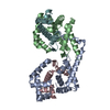
- PDB-6nzf: CRYSTAL STRUCTURE OF TYROSINE KINASE 2 JH2 (PSEUDO KINASE DOMAIN)... -
+ Open data

- Basic information
| Entry | Database: PDB / ID: 6nzf | ||||||
|---|---|---|---|---|---|---|---|
| Title | CRYSTAL STRUCTURE OF TYROSINE KINASE 2 JH2 (PSEUDO KINASE DOMAIN) COMPLEXED WITH Compound_5 AKA 4-[(2-CARBAMOYLPHEN YL)AMINO]-6-[(5-FLUOROPYRIDIN-2-YL)AMINO]-N-METHYLPYRIDINE -3-CARBOXAMIDE | ||||||
 Components Components | Non-receptor tyrosine-protein kinase TYK2 | ||||||
 Keywords Keywords | TRANSFERASE / JTK1 | ||||||
| Function / homology |  Function and homology information Function and homology informationtype III interferon-mediated signaling pathway / interleukin-10-mediated signaling pathway / interleukin-12 receptor complex / interleukin-23 receptor complex / Interleukin-23 signaling / interleukin-23-mediated signaling pathway / positive regulation of T-helper 17 type immune response / interleukin-12-mediated signaling pathway / positive regulation of NK T cell proliferation / Interleukin-12 signaling ...type III interferon-mediated signaling pathway / interleukin-10-mediated signaling pathway / interleukin-12 receptor complex / interleukin-23 receptor complex / Interleukin-23 signaling / interleukin-23-mediated signaling pathway / positive regulation of T-helper 17 type immune response / interleukin-12-mediated signaling pathway / positive regulation of NK T cell proliferation / Interleukin-12 signaling / IL-6-type cytokine receptor ligand interactions / Interleukin-27 signaling / Interleukin-35 Signalling / growth hormone receptor binding / Other interleukin signaling / extrinsic component of plasma membrane / Interleukin-20 family signaling / extrinsic component of cytoplasmic side of plasma membrane / Interleukin-6 signaling / type I interferon-mediated signaling pathway / MAPK3 (ERK1) activation / MAPK1 (ERK2) activation / Interleukin-10 signaling / positive regulation of interleukin-17 production / Regulation of IFNA/IFNB signaling / positive regulation of natural killer cell proliferation / growth hormone receptor signaling pathway via JAK-STAT / type II interferon-mediated signaling pathway / cell surface receptor signaling pathway via JAK-STAT / Signaling by CSF3 (G-CSF) / positive regulation of T cell proliferation / positive regulation of receptor signaling pathway via JAK-STAT / non-specific protein-tyrosine kinase / non-membrane spanning protein tyrosine kinase activity / cellular response to virus / Inactivation of CSF3 (G-CSF) signaling / positive regulation of protein localization to nucleus / Evasion by RSV of host interferon responses / positive regulation of type II interferon production / cytoplasmic side of plasma membrane / cytokine-mediated signaling pathway / Interferon alpha/beta signaling / Signaling by ALK fusions and activated point mutants / protein tyrosine kinase activity / Interleukin-4 and Interleukin-13 signaling / Potential therapeutics for SARS / protein phosphorylation / cell differentiation / signaling receptor complex / cell population proliferation / intracellular signal transduction / immune response / SARS-CoV-2 activates/modulates innate and adaptive immune responses / extracellular exosome / ATP binding / nucleus / plasma membrane / cytoplasm / cytosol Similarity search - Function | ||||||
| Biological species |  Homo sapiens (human) Homo sapiens (human) | ||||||
| Method |  X-RAY DIFFRACTION / SYNCHROTRON / MOLECULAR REPLACEMENT / Resolution: 2.39 Å X-RAY DIFFRACTION / SYNCHROTRON / MOLECULAR REPLACEMENT / Resolution: 2.39 Å | ||||||
 Authors Authors | Muckelbauer, J.M. | ||||||
 Citation Citation | Journal: J.Med.Chem. / Year: 2019 Title: Identification ofN-Methyl Nicotinamide andN-Methyl Pyridazine-3-Carboxamide Pseudokinase Domain Ligands as Highly Selective Allosteric Inhibitors of Tyrosine Kinase 2 (TYK2). Authors: Moslin, R. / Zhang, Y. / Wrobleski, S.T. / Lin, S. / Mertzman, M. / Spergel, S. / Tokarski, J.S. / Strnad, J. / Gillooly, K. / McIntyre, K.W. / Zupa-Fernandez, A. / Cheng, L. / Sun, H. / ...Authors: Moslin, R. / Zhang, Y. / Wrobleski, S.T. / Lin, S. / Mertzman, M. / Spergel, S. / Tokarski, J.S. / Strnad, J. / Gillooly, K. / McIntyre, K.W. / Zupa-Fernandez, A. / Cheng, L. / Sun, H. / Chaudhry, C. / Huang, C. / D'Arienzo, C. / Heimrich, E. / Yang, X. / Muckelbauer, J.K. / Chang, C. / Tredup, J. / Mulligan, D. / Xie, D. / Aranibar, N. / Chiney, M. / Burke, J.R. / Lombardo, L. / Carter, P.H. / Weinstein, D.S. | ||||||
| History |
|
- Structure visualization
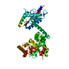
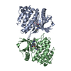
| Structure viewer | Molecule: MolmilJmol/JSmol |
|---|
- Downloads & links
-Download
| PDBx/mmCIF format | 6nzf.cif.gz | 113.8 KB | Display | PDBx/mmCIF format |
|---|---|---|---|---|
| PDB format | pdb6nzf.ent.gz | 84 KB | Display | PDB format |
| PDBx/mmJSON format | 6nzf.json.gz | Tree view | PDBx/mmJSON format | |
| Others |  Other downloads Other downloads |
-Validation report
| Arichive directory | https://data.pdbj.org/pub/pdb/validation_reports/nz/6nzfftp://data.pdbj.org/pub/pdb/validation_reports/nz/6nzf | HTTPS FTP |
|---|
-Related structure data
| Related structure data | 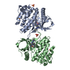 6nzeC 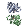 6nzhC 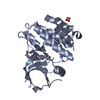 4wovS S: Starting model for refinement C: citing same article ( |
|---|---|
| Similar structure data |

-Links
 PDBj
PDBj


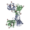
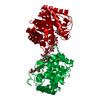
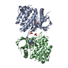
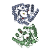
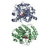
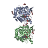
- Assembly
| Deposited unit | 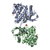
| ||||||||
|---|---|---|---|---|---|---|---|---|---|
| 1 |
| ||||||||
| Unit cell |
|
-Components
| #1: Protein | Mass: 35499.156 Da / Num. of mol.: 2 / Fragment: Pseudo kinase domain, residues 575-869 Source method: isolated from a genetically manipulated source Source: (gene. exp.) Homo sapiens (human) / Gene: TYK2 / Plasmid: pFastBac1 / Production host:   Spodoptera frugiperda (fall armyworm) / Strain (production host): SF9 Spodoptera frugiperda (fall armyworm) / Strain (production host): SF9References: UniProt: P29597, non-specific protein-tyrosine kinase #2: Chemical | 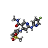  Mass: 415.441 Da / Num. of mol.: 2 / Source method: obtained synthetically / Formula: C19H18FN5O3S Mass: 415.441 Da / Num. of mol.: 2 / Source method: obtained synthetically / Formula: C19H18FN5O3S#3: Chemical |   Mass: 96.063 Da / Num. of mol.: 3 / Source method: obtained synthetically / Formula: SO4 Mass: 96.063 Da / Num. of mol.: 3 / Source method: obtained synthetically / Formula: SO4#4: Water | ChemComp-HOH / |  Mass: 18.015 Da / Num. of mol.: 34 / Source method: isolated from a natural source / Formula: H2O Mass: 18.015 Da / Num. of mol.: 34 / Source method: isolated from a natural source / Formula: H2O |
|---|
-Experimental details
-Experiment
| Experiment | Method: X-RAY DIFFRACTION / Number of used crystals: 1 |
|---|
- Sample preparation
| Crystal | Density Matthews: 2.27 Å3/Da / Density % sol: 45.79 % |
|---|---|
| Crystal grow | Temperature: 293 K / Method: vapor diffusion, hanging drop / pH: 7.5 Details: 200 mM ammonium sulfate, and 100 mM HEPES buffer, pH 7.5, 30%(W/V) PEG 5000 (Methyl Ether) |
-Data collection
| Diffraction | Mean temperature: 100 K / Serial crystal experiment: N |
|---|---|
| Diffraction source | Source: SYNCHROTRON / Site: APS  / Beamline: 17-ID / Wavelength: 1 Å / Beamline: 17-ID / Wavelength: 1 Å |
| Detector | Type: DECTRIS PILATUS 6M / Detector: PIXEL / Date: Jun 13, 2012 |
| Radiation | Protocol: SINGLE WAVELENGTH / Monochromatic (M) / Laue (L): M / Scattering type: x-ray |
| Radiation wavelength | Wavelength: 1 Å / Relative weight: 1 |
| Reflection | Resolution: 2.39→46.29 Å / Num. obs: 25218 / % possible obs: 99.2 % / Redundancy: 3.3 % / Biso Wilson estimate: 56.73 Å2 / Rsym value: 0.054 / Net I/σ(I): 13.4 |
| Reflection shell | Resolution: 7.55→46.29 Å / Redundancy: 3.1 % / Mean I/σ(I) obs: 39.3 / Rsym value: 0.02 / % possible all: 97.3 |
- Processing
| Software |
| ||||||||||||||||||||||||||||||||||||||||||||||||||||||||||||||||||||||||||||||||||||||||||||||||||||||||||||
|---|---|---|---|---|---|---|---|---|---|---|---|---|---|---|---|---|---|---|---|---|---|---|---|---|---|---|---|---|---|---|---|---|---|---|---|---|---|---|---|---|---|---|---|---|---|---|---|---|---|---|---|---|---|---|---|---|---|---|---|---|---|---|---|---|---|---|---|---|---|---|---|---|---|---|---|---|---|---|---|---|---|---|---|---|---|---|---|---|---|---|---|---|---|---|---|---|---|---|---|---|---|---|---|---|---|---|---|---|---|
| Refinement | Method to determine structure: MOLECULAR REPLACEMENT Starting model: 4WOV Resolution: 2.39→46.29 Å / Cor.coef. Fo:Fc: 0.915 / Cor.coef. Fo:Fc free: 0.899 / Rfactor Rfree error: 0 / SU R Cruickshank DPI: 0.301 / Cross valid method: THROUGHOUT / σ(F): 0 / SU R Blow DPI: 0.283 / SU Rfree Blow DPI: 0.207 / SU Rfree Cruickshank DPI: 0.214
| ||||||||||||||||||||||||||||||||||||||||||||||||||||||||||||||||||||||||||||||||||||||||||||||||||||||||||||
| Displacement parameters | Biso max: 125.85 Å2 / Biso mean: 51.05 Å2 / Biso min: 30.23 Å2
| ||||||||||||||||||||||||||||||||||||||||||||||||||||||||||||||||||||||||||||||||||||||||||||||||||||||||||||
| Refine analyze | Luzzati coordinate error obs: 0.33 Å | ||||||||||||||||||||||||||||||||||||||||||||||||||||||||||||||||||||||||||||||||||||||||||||||||||||||||||||
| Refinement step | Cycle: final / Resolution: 2.39→46.29 Å
| ||||||||||||||||||||||||||||||||||||||||||||||||||||||||||||||||||||||||||||||||||||||||||||||||||||||||||||
| Refine LS restraints |
| ||||||||||||||||||||||||||||||||||||||||||||||||||||||||||||||||||||||||||||||||||||||||||||||||||||||||||||
| LS refinement shell | Resolution: 2.39→2.49 Å / Rfactor Rfree error: 0 / Total num. of bins used: 13
|
