 Movie
Movie Controller


+ Open data

- Basic information
| Entry | Database: PDB / ID: 6nxc | ||||||
|---|---|---|---|---|---|---|---|
| Title | ECAI(T162A) MUTANT IN COMPLEX WITH CITRATE AT PH 4 | ||||||
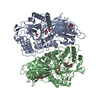
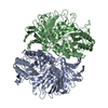
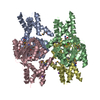
 Components Components | L-asparaginase 1 | ||||||
 Keywords Keywords | HYDROLASE / hydrolysis of L-asparagine | ||||||
| Function / homology |  Function and homology information Function and homology information: / asparaginase / asparaginase activity / protein homotetramerization / identical protein binding / cytosol / cytoplasm Similarity search - Function | ||||||
| Biological species |  | ||||||
| Method |  X-RAY DIFFRACTION / SYNCHROTRON / Resolution: 1.74 Å X-RAY DIFFRACTION / SYNCHROTRON / Resolution: 1.74 Å | ||||||
 Authors Authors | Lubkowski, J. / Wlodawer, A. | ||||||
 Citation Citation | Journal: J. Mol. Biol. / Year: 2007 Title: Crystal structure and allosteric regulation of the cytoplasmic Escherichia coli L-asparaginase I. Authors: Yun, M.K. / Nourse, A. / White, S.W. / Rock, C.O. / Heath, R.J. | ||||||
| History |
| ||||||
| Remark 0 | THIS ENTRY 6NXC REFLECTS AN ALTERNATIVE MODELING OF THE STRUCTURAL DATA IN R2HIMSF ORIGINAL DATA ...THIS ENTRY 6NXC REFLECTS AN ALTERNATIVE MODELING OF THE STRUCTURAL DATA IN R2HIMSF ORIGINAL DATA DETERMINED BY AUTHOR: M.K.YUN,A.NOURSE,S.W.WHITE,C.O.ROCK,R.J.HEATH. |
- Structure visualization
| Structure viewer | Molecule: MolmilJmol/JSmol |
|---|
- Downloads & links
-Download
| PDBx/mmCIF format | 6nxc.cif.gz | 280.7 KB | Display | PDBx/mmCIF format |
|---|---|---|---|---|
| PDB format | pdb6nxc.ent.gz | 222.5 KB | Display | PDB format |
| PDBx/mmJSON format | 6nxc.json.gz | Tree view | PDBx/mmJSON format | |
| Others |  Other downloads Other downloads |
-Validation report
| Arichive directory | https://data.pdbj.org/pub/pdb/validation_reports/nx/6nxcftp://data.pdbj.org/pub/pdb/validation_reports/nx/6nxc | HTTPS FTP |
|---|
-Related structure data
| Related structure data |  6nx6C  6nx7C  6nx8C  6nx9C  6nxaC 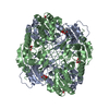 6nxbC  6nxdC C: citing same article ( |
|---|---|
| Similar structure data |


-Links
 PDBj
PDBj- Assembly
| Deposited unit | 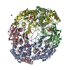
| ||||||||
|---|---|---|---|---|---|---|---|---|---|
| 1 |
| ||||||||
| Unit cell |
|
-Components
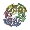
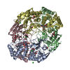
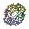
-Protein , 1 types, 4 molecules ABCD
| #1: Protein | Mass: 39190.348 Da / Num. of mol.: 4 / Mutation: T162A Source method: isolated from a genetically manipulated source Source: (gene. exp.) Strain: K12 / Gene: ansA, b1767, JW1756 / Plasmid: pET-15b / Production host: |
|---|
-Non-polymers , 6 types, 595 molecules 


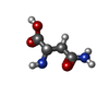

| #2: Chemical | ChemComp-CIT /  Mass: 192.124 Da / Num. of mol.: 4 / Source method: obtained synthetically / Formula: C6H8O7 / Feature type: SUBJECT OF INVESTIGATION Mass: 192.124 Da / Num. of mol.: 4 / Source method: obtained synthetically / Formula: C6H8O7 / Feature type: SUBJECT OF INVESTIGATION#3: Chemical | ChemComp-EDO /  Mass: 62.068 Da / Num. of mol.: 11 / Source method: obtained synthetically / Formula: C2H6O2 Mass: 62.068 Da / Num. of mol.: 11 / Source method: obtained synthetically / Formula: C2H6O2#4: Chemical | ChemComp-CL /  Mass: 35.453 Da / Num. of mol.: 10 / Source method: obtained synthetically / Formula: Cl Mass: 35.453 Da / Num. of mol.: 10 / Source method: obtained synthetically / Formula: Cl#5: Chemical | ChemComp-GOL / |  Mass: 92.094 Da / Num. of mol.: 1 / Source method: obtained synthetically / Formula: C3H8O3 Mass: 92.094 Da / Num. of mol.: 1 / Source method: obtained synthetically / Formula: C3H8O3#6: Chemical | ChemComp-ASN /  Type: L-peptide linking / Mass: 132.118 Da / Num. of mol.: 4 / Source method: obtained synthetically / Formula: C4H8N2O3 / Feature type: SUBJECT OF INVESTIGATION Type: L-peptide linking / Mass: 132.118 Da / Num. of mol.: 4 / Source method: obtained synthetically / Formula: C4H8N2O3 / Feature type: SUBJECT OF INVESTIGATION#7: Water | ChemComp-HOH / | Mass: 18.015 Da / Num. of mol.: 565 / Source method: isolated from a natural source / Formula: H2O |
|---|
-Details
| Has protein modification | Y |
|---|
-Experimental details
-Experiment
| Experiment | Method: X-RAY DIFFRACTION / Number of used crystals: 1 |
|---|
- Sample preparation
| Crystal | Density Matthews: 2.15 Å3/Da / Density % sol: 42.67 % / Description: AUTHOR USED THE SF DATA FROM ENTRY 2HIM |
|---|---|
| Crystal grow | Temperature: 291 K / Method: vapor diffusion, hanging drop / pH: 4 Details: Citrate buffer, Sodium Chloride, pH 4.0, VAPOR DIFFUSION, HANGING DROP, temperature 291K |
-Data collection
| Diffraction | Mean temperature: 100 K / Serial crystal experiment: N |
|---|---|
| Diffraction source | Source: SYNCHROTRON / Site: APS  / Beamline: 22-BM / Wavelength: 0.97923 Å / Beamline: 22-BM / Wavelength: 0.97923 Å |
| Detector | Type: MARRESEARCH / Detector: CCD / Date: Feb 28, 2006 |
| Radiation | Protocol: SINGLE WAVELENGTH / Monochromatic (M) / Laue (L): M / Scattering type: x-ray |
| Radiation wavelength | Wavelength: 0.97923 Å / Relative weight: 1 |
| Reflection | Resolution: 1.74→50 Å / Num. obs: 132353 / % possible obs: 98.3 % / Observed criterion σ(I): -3 / Redundancy: 3 % / Net I/σ(I): 14.8 |
| Reflection shell | Resolution: 1.74→1.81 Å |
- Processing
| Software |
| ||||||||||||||||||||||||||||||||||||||||||||||||||||||||||||
|---|---|---|---|---|---|---|---|---|---|---|---|---|---|---|---|---|---|---|---|---|---|---|---|---|---|---|---|---|---|---|---|---|---|---|---|---|---|---|---|---|---|---|---|---|---|---|---|---|---|---|---|---|---|---|---|---|---|---|---|---|---|
| Refinement | Resolution: 1.74→46.54 Å / Cor.coef. Fo:Fc: 0.961 / Cor.coef. Fo:Fc free: 0.946 / SU B: 2.236 / SU ML: 0.072 / SU R Cruickshank DPI: 0.113 / Cross valid method: THROUGHOUT / σ(F): 0 / ESU R: 0.113 / ESU R Free: 0.113 Details: HYDROGENS HAVE BEEN ADDED IN THE RIDING POSITIONS U VALUES : REFINED INDIVIDUALLY
| ||||||||||||||||||||||||||||||||||||||||||||||||||||||||||||
| Solvent computation | Ion probe radii: 0.8 Å / Shrinkage radii: 0.8 Å / VDW probe radii: 1.2 Å | ||||||||||||||||||||||||||||||||||||||||||||||||||||||||||||
| Displacement parameters | Biso max: 133.73 Å2 / Biso mean: 25.698 Å2 / Biso min: 11.57 Å2
| ||||||||||||||||||||||||||||||||||||||||||||||||||||||||||||
| Refinement step | Cycle: final / Resolution: 1.74→46.54 Å
| ||||||||||||||||||||||||||||||||||||||||||||||||||||||||||||
| Refine LS restraints |
| ||||||||||||||||||||||||||||||||||||||||||||||||||||||||||||
| LS refinement shell | Resolution: 1.744→1.79 Å / Rfactor Rfree error: 0 / Total num. of bins used: 20
|
