Entry Database : PDB / ID : 6nr7Title Rerefinement of chicken vinculin Vinculin Keywords / / / / Function / homology Function Domain/homology Component
/ / / / / / / / / / / / / / / / / / / / / / / / / / / / / / / / / / / / / / / / / / / / / / / / / / / / / / / / / / / / / / / / / / / / / / / / / / / / / / / / / / Biological species Gallus gallus (chicken)Method / / / Resolution : 3 Å Authors Stec, B. Journal : To be published Title : Refined model of chicken vinculin suggests the mechanism of activation by helical super-bundle unfurlingAuthors : Stec, D.L. / Stec, B. History Deposition Jan 23, 2019 Deposition site / Processing site Revision 1.0 Jan 29, 2020 Provider / Type Revision 1.1 Oct 11, 2023 Group / Database references / Refinement descriptionCategory chem_comp_atom / chem_comp_bond ... chem_comp_atom / chem_comp_bond / database_2 / pdbx_initial_refinement_model Item / _database_2.pdbx_database_accession
Show all Show less
 Movie
Movie Controller


 Open data
 Basic information
 Components
 Keywords
 Function and homology information
 X-RAY DIFFRACTION /
  Authors
 Citation
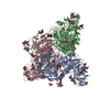
 Structure visualization
 Downloads & links
 Other downloads
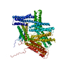

 PDBj
PDBj
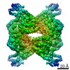
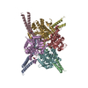
 Assembly
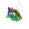


 Mass: 96.063 Da / Num. of mol.: 2 / Source method: obtained synthetically / Formula: SO4
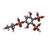
 Mass: 494.174 Da / Num. of mol.: 1 / Source method: obtained synthetically / Formula: C9H21O17P3
 Mass: 94.971 Da / Num. of mol.: 2 / Source method: obtained synthetically / Formula: PO4
 Mass: 18.015 Da / Num. of mol.: 58 / Source method: isolated from a natural source / Formula: H2O
 Sample preparation
 / Beamline: BL9-1 / Wavelength: 0.97955 Å
 Processing