 Movie
Movie Controller


+ Open data

- Basic information
| Entry | Database: PDB / ID: 6nqz | ||||||
|---|---|---|---|---|---|---|---|
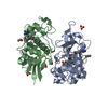
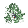
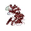
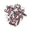
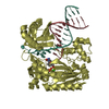
| Title | Flagellar protein FcpB from Leptospira interrogans | ||||||
 Components Components | Flagellar coiling protein B | ||||||
 Keywords Keywords | PROTEIN FIBRIL / flagellar filament | ||||||
| Function / homology | Chem-I3C / IODIDE ION / Uncharacterized protein Function and homology information Function and homology information | ||||||
| Biological species |  Leptospira interrogans serogroup Icterohaemorrhagiae serovar copenhageni (bacteria) Leptospira interrogans serogroup Icterohaemorrhagiae serovar copenhageni (bacteria) | ||||||
| Method |  X-RAY DIFFRACTION / SAD / Resolution: 2.58 Å X-RAY DIFFRACTION / SAD / Resolution: 2.58 Å | ||||||
 Authors Authors | Trajtenberg, F. / Larrieux, N. / Buschiazzo, A. | ||||||
 Citation Citation | Journal: Elife / Year: 2020 Title: An asymmetric sheath controls flagellar supercoiling and motility in the Leptospira spirochete. Authors: Gibson, K. / Trajtenberg, F. / Brady, M. / San Martin, F. / Mechaly, A. / Wunder, E. / Picardeau, M. / Ko, A. / Buschiazzo, A. / Sindelar, C. | ||||||
| History |
|
- Structure visualization
| Structure viewer | Molecule: MolmilJmol/JSmol |
|---|
- Downloads & links
-Download
| PDBx/mmCIF format | 6nqz.cif.gz | 101.2 KB | Display | PDBx/mmCIF format |
|---|---|---|---|---|
| PDB format | pdb6nqz.ent.gz | 76.2 KB | Display | PDB format |
| PDBx/mmJSON format | 6nqz.json.gz | Tree view | PDBx/mmJSON format | |
| Others |  Other downloads Other downloads |
-Validation report
| Arichive directory | https://data.pdbj.org/pub/pdb/validation_reports/nq/6nqzftp://data.pdbj.org/pub/pdb/validation_reports/nq/6nqz | HTTPS FTP |
|---|
-Related structure data


-Links
 PDBj
PDBj- Assembly
| Deposited unit | 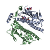
| ||||||||
|---|---|---|---|---|---|---|---|---|---|
| 1 |
| ||||||||
| Unit cell |
|
-Components
| #1: Protein | Mass: 30177.816 Da / Num. of mol.: 2 Source method: isolated from a genetically manipulated source Source: (gene. exp.) Leptospira interrogans serogroup Icterohaemorrhagiae serovar copenhageni (strain Fiocruz L1-130) (bacteria)Strain: Fiocruz L1-130 / Gene: LIC_11848 / Production host: #2: Chemical | ChemComp-I3C / |   Mass: 558.835 Da / Num. of mol.: 1 / Source method: obtained synthetically / Formula: C8H4I3NO4 Mass: 558.835 Da / Num. of mol.: 1 / Source method: obtained synthetically / Formula: C8H4I3NO4#3: Chemical |   Mass: 126.904 Da / Num. of mol.: 3 / Source method: obtained synthetically / Formula: I Mass: 126.904 Da / Num. of mol.: 3 / Source method: obtained synthetically / Formula: I#4: Chemical |   Mass: 92.094 Da / Num. of mol.: 2 / Source method: obtained synthetically / Formula: C3H8O3 Mass: 92.094 Da / Num. of mol.: 2 / Source method: obtained synthetically / Formula: C3H8O3#5: Water | ChemComp-HOH / |  Mass: 18.015 Da / Num. of mol.: 31 / Source method: isolated from a natural source / Formula: H2O Mass: 18.015 Da / Num. of mol.: 31 / Source method: isolated from a natural source / Formula: H2OHas protein modification | Y | |
|---|
-Experimental details
-Experiment
| Experiment | Method: X-RAY DIFFRACTION / Number of used crystals: 1 |
|---|
- Sample preparation
| Crystal | Density Matthews: 2.22 Å3/Da / Density % sol: 44.59 % |
|---|---|
| Crystal grow | Temperature: 293 K / Method: vapor diffusion, hanging drop / pH: 6.5 / Details: (NH4)I, PEG 3350, MES, Glycerol |
-Data collection
| Diffraction | Mean temperature: 109 K / Serial crystal experiment: N | |||||||||||||||||||||||||||||||||||||||||||||||||||||||||||||||||||||||||||||||||||||||||||||||||||
|---|---|---|---|---|---|---|---|---|---|---|---|---|---|---|---|---|---|---|---|---|---|---|---|---|---|---|---|---|---|---|---|---|---|---|---|---|---|---|---|---|---|---|---|---|---|---|---|---|---|---|---|---|---|---|---|---|---|---|---|---|---|---|---|---|---|---|---|---|---|---|---|---|---|---|---|---|---|---|---|---|---|---|---|---|---|---|---|---|---|---|---|---|---|---|---|---|---|---|---|---|
| Diffraction source | Source: ROTATING ANODE / Type: RIGAKU MICROMAX-007 HF / Wavelength: 1.54179 Å | |||||||||||||||||||||||||||||||||||||||||||||||||||||||||||||||||||||||||||||||||||||||||||||||||||
| Detector | Type: MAR scanner 345 mm plate / Detector: IMAGE PLATE / Date: Apr 28, 2012 | |||||||||||||||||||||||||||||||||||||||||||||||||||||||||||||||||||||||||||||||||||||||||||||||||||
| Radiation | Protocol: SINGLE WAVELENGTH / Monochromatic (M) / Laue (L): M / Scattering type: x-ray | |||||||||||||||||||||||||||||||||||||||||||||||||||||||||||||||||||||||||||||||||||||||||||||||||||
| Radiation wavelength | Wavelength: 1.54179 Å / Relative weight: 1 | |||||||||||||||||||||||||||||||||||||||||||||||||||||||||||||||||||||||||||||||||||||||||||||||||||
| Reflection | Resolution: 2.577→67.232 Å / Num. obs: 17479 / % possible obs: 99.2 % / Redundancy: 3.6 % / Biso Wilson estimate: 42.84 Å2 / Rpim(I) all: 0.078 / Rrim(I) all: 0.15 / Rsym value: 0.128 / Net I/av σ(I): 5.8 / Net I/σ(I): 8.1 | |||||||||||||||||||||||||||||||||||||||||||||||||||||||||||||||||||||||||||||||||||||||||||||||||||
| Reflection shell | Diffraction-ID: 1
|
- Processing
| Software |
| ||||||||||||||||||||||||||||||||||||||||||||||||||||||||||||||||||||||||||||||||||||||||||||||||||||||||||||
|---|---|---|---|---|---|---|---|---|---|---|---|---|---|---|---|---|---|---|---|---|---|---|---|---|---|---|---|---|---|---|---|---|---|---|---|---|---|---|---|---|---|---|---|---|---|---|---|---|---|---|---|---|---|---|---|---|---|---|---|---|---|---|---|---|---|---|---|---|---|---|---|---|---|---|---|---|---|---|---|---|---|---|---|---|---|---|---|---|---|---|---|---|---|---|---|---|---|---|---|---|---|---|---|---|---|---|---|---|---|
| Refinement | Method to determine structure: SAD / Resolution: 2.58→37.01 Å / Cor.coef. Fo:Fc: 0.907 / Cor.coef. Fo:Fc free: 0.856 / SU R Cruickshank DPI: 0.529 / Cross valid method: THROUGHOUT / σ(F): 0 / SU R Blow DPI: 0.539 / SU Rfree Blow DPI: 0.286 / SU Rfree Cruickshank DPI: 0.289
| ||||||||||||||||||||||||||||||||||||||||||||||||||||||||||||||||||||||||||||||||||||||||||||||||||||||||||||
| Displacement parameters | Biso max: 122.31 Å2 / Biso mean: 33.32 Å2 / Biso min: 3.06 Å2
| ||||||||||||||||||||||||||||||||||||||||||||||||||||||||||||||||||||||||||||||||||||||||||||||||||||||||||||
| Refine analyze | Luzzati coordinate error obs: 0.35 Å | ||||||||||||||||||||||||||||||||||||||||||||||||||||||||||||||||||||||||||||||||||||||||||||||||||||||||||||
| Refinement step | Cycle: final / Resolution: 2.58→37.01 Å
| ||||||||||||||||||||||||||||||||||||||||||||||||||||||||||||||||||||||||||||||||||||||||||||||||||||||||||||
| Refine LS restraints |
| ||||||||||||||||||||||||||||||||||||||||||||||||||||||||||||||||||||||||||||||||||||||||||||||||||||||||||||
| LS refinement shell | Resolution: 2.58→2.6 Å / Rfactor Rfree error: 0 / Total num. of bins used: 50
|
