 Movie
Movie Controller

[English] 日本語
 Yorodumi
- PDB-6nqg: Xanthomonas citri Phospho-PGM in complex with glucopyranosyl-1-me... -
+ Open data

- Basic information
| Entry | Database: PDB / ID: 6nqg | ||||||
|---|---|---|---|---|---|---|---|
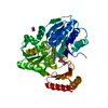
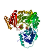
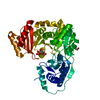
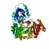
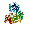
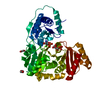
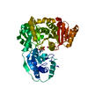
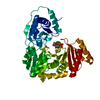
| Title | Xanthomonas citri Phospho-PGM in complex with glucopyranosyl-1-methyl-phosphonic acid at room temperature | ||||||
 Components Components | Phosphoglucomutase | ||||||
 Keywords Keywords | ISOMERASE / phosphoglucomutase | ||||||
| Function / homology |  Function and homology information Function and homology informationintramolecular phosphotransferase activity / carbohydrate metabolic process / magnesium ion binding Similarity search - Function | ||||||
| Biological species |  Xanthomonas axonopodis pv. citri (bacteria) Xanthomonas axonopodis pv. citri (bacteria) | ||||||
| Method |  X-RAY DIFFRACTION / SYNCHROTRON / MOLECULAR REPLACEMENT / Resolution: 2.05 Å X-RAY DIFFRACTION / SYNCHROTRON / MOLECULAR REPLACEMENT / Resolution: 2.05 Å | ||||||
 Authors Authors | Stiers, K.M. / Beamer, L.J. | ||||||
| Funding support |  United States, 1items United States, 1items
| ||||||
 Citation Citation | Journal: Struct Dyn. / Year: 2019 Title: Structural and dynamical description of the enzymatic reaction of a phosphohexomutase. Authors: Stiers, K.M. / Graham, A.C. / Zhu, J.S. / Jakeman, D.L. / Nix, J.C. / Beamer, L.J. | ||||||
| History |
|
- Structure visualization
| Structure viewer | Molecule: MolmilJmol/JSmol |
|---|
- Downloads & links
-Download
| PDBx/mmCIF format | 6nqg.cif.gz | 195.8 KB | Display | PDBx/mmCIF format |
|---|---|---|---|---|
| PDB format | pdb6nqg.ent.gz | 154.8 KB | Display | PDB format |
| PDBx/mmJSON format | 6nqg.json.gz | Tree view | PDBx/mmJSON format | |
| Others |  Other downloads Other downloads |
-Validation report
| Arichive directory | https://data.pdbj.org/pub/pdb/validation_reports/nq/6nqgftp://data.pdbj.org/pub/pdb/validation_reports/nq/6nqg | HTTPS FTP |
|---|
-Related structure data
| Related structure data | 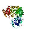 6nn1C 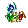 6nn2C  6nnnC  6nnoC  6nnpC 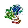 6nnsC  6nntC  6nnuC  6nolC  6noqC  6np8C  6npxC 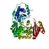 6nqeC 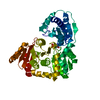 6nqfC 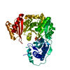 5bmnS C: citing same article ( S: Starting model for refinement |
|---|---|
| Similar structure data |


-Links
 PDBj
PDBj- Assembly
| Deposited unit | 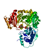
| ||||||||
|---|---|---|---|---|---|---|---|---|---|
| 1 |
| ||||||||
| Unit cell |
|
-Components
| #1: Protein | Mass: 51431.621 Da / Num. of mol.: 1 Source method: isolated from a genetically manipulated source Source: (gene. exp.) Xanthomonas axonopodis pv. citri (strain 306) (bacteria)Strain: 306 / Gene: xanA, XAC3579 / Production host: |
|---|---|
| #2: Chemical | ChemComp-CA /   Mass: 40.078 Da / Num. of mol.: 1 / Source method: obtained synthetically / Formula: Ca Mass: 40.078 Da / Num. of mol.: 1 / Source method: obtained synthetically / Formula: Ca |
| #3: Sugar | ChemComp-GPM / (  Type: D-saccharide / Mass: 258.163 Da / Num. of mol.: 1 Type: D-saccharide / Mass: 258.163 Da / Num. of mol.: 1Source method: isolated from a genetically manipulated source Formula: C7H15O8P |
| #4: Water | ChemComp-HOH /  Mass: 18.015 Da / Num. of mol.: 122 / Source method: isolated from a natural source / Formula: H2O Mass: 18.015 Da / Num. of mol.: 122 / Source method: isolated from a natural source / Formula: H2O |
| Has protein modification | Y |
-Experimental details
-Experiment
| Experiment | Method: X-RAY DIFFRACTION / Number of used crystals: 1 |
|---|
- Sample preparation
| Crystal | Density Matthews: 2.05 Å3/Da / Density % sol: 39.93 % |
|---|---|
| Crystal grow | Temperature: 277 K / Method: vapor diffusion, hanging drop / pH: 7.5 / Details: 22% PEG 8000, 0.2M MgCl, 0.1M HEPES |
-Data collection
| Diffraction | Mean temperature: 298 K / Serial crystal experiment: N |
|---|---|
| Diffraction source | Source: SYNCHROTRON / Site: ALS / Beamline: 4.2.2 / Wavelength: 1.00003 Å |
| Detector | Type: RDI CMOS_8M / Detector: CMOS / Date: Mar 28, 2018 |
| Radiation | Protocol: SINGLE WAVELENGTH / Monochromatic (M) / Laue (L): M / Scattering type: x-ray |
| Radiation wavelength | Wavelength: 1.00003 Å / Relative weight: 1 |
| Reflection | Resolution: 2.05→52.151 Å / Num. obs: 27455 / % possible obs: 100 % / Redundancy: 13.8 % / CC1/2: 0.989 / Rmerge(I) obs: 0.482 / Rpim(I) all: 0.133 / Rrim(I) all: 0.5 / Net I/σ(I): 6.5 |
| Reflection shell | Resolution: 2.05→2.11 Å / Redundancy: 12.3 % / Rmerge(I) obs: 5.633 / Mean I/σ(I) obs: 0.6 / Num. unique obs: 2105 / CC1/2: 0.301 / Rpim(I) all: 1.661 / Rrim(I) all: 5.88 / % possible all: 100 |
- Processing
| Software |
| |||||||||||||||||||||||||||||||||||||||||||||||||||||||||||||||||||||||||||||
|---|---|---|---|---|---|---|---|---|---|---|---|---|---|---|---|---|---|---|---|---|---|---|---|---|---|---|---|---|---|---|---|---|---|---|---|---|---|---|---|---|---|---|---|---|---|---|---|---|---|---|---|---|---|---|---|---|---|---|---|---|---|---|---|---|---|---|---|---|---|---|---|---|---|---|---|---|---|---|
| Refinement | Method to determine structure: MOLECULAR REPLACEMENT Starting model: 5BMN Resolution: 2.05→52.151 Å / SU ML: 0.39 / Cross valid method: FREE R-VALUE / σ(F): 0.03 / Phase error: 26.01
| |||||||||||||||||||||||||||||||||||||||||||||||||||||||||||||||||||||||||||||
| Solvent computation | Shrinkage radii: 0.9 Å / VDW probe radii: 1.11 Å | |||||||||||||||||||||||||||||||||||||||||||||||||||||||||||||||||||||||||||||
| Refinement step | Cycle: LAST / Resolution: 2.05→52.151 Å
| |||||||||||||||||||||||||||||||||||||||||||||||||||||||||||||||||||||||||||||
| Refine LS restraints |
| |||||||||||||||||||||||||||||||||||||||||||||||||||||||||||||||||||||||||||||
| LS refinement shell |
| |||||||||||||||||||||||||||||||||||||||||||||||||||||||||||||||||||||||||||||
| Refinement TLS params. | Method: refined / Refine-ID: X-RAY DIFFRACTION
| |||||||||||||||||||||||||||||||||||||||||||||||||||||||||||||||||||||||||||||
| Refinement TLS group |
|
