 Movie
Movie Controller


+ Open data

- Basic information
| Entry | Database: PDB / ID: 6nqc | ||||||
|---|---|---|---|---|---|---|---|
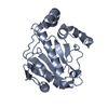
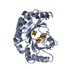
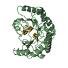
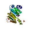
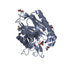
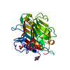
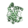
| Title | Crystal structure of a peptidase from an acI-B1 Actinobacterium | ||||||
 Components Components | Cyanophycinase-like exopeptidase | ||||||
 Keywords Keywords | HYDROLASE / Peptidase | ||||||
| Function / homology | Peptidase S51 / Peptidase family S51 / Class I glutamine amidotransferase-like / serine-type peptidase activity / Cysnophycinase-like exopeptidase Function and homology information Function and homology information | ||||||
| Biological species |  actinobacterium SCGC AAA027-L06 (bacteria) actinobacterium SCGC AAA027-L06 (bacteria) | ||||||
| Method |  X-RAY DIFFRACTION / SYNCHROTRON / MOLECULAR REPLACEMENT / Resolution: 1.94 Å X-RAY DIFFRACTION / SYNCHROTRON / MOLECULAR REPLACEMENT / Resolution: 1.94 Å | ||||||
 Authors Authors | Forest, K.T. / Dwulit-Smith, J.R. / Satyshur, K.A. | ||||||
| Funding support |  United States, 1items United States, 1items
| ||||||
 Citation Citation | Journal: To Be Published Title: Structure of a peptidase from an acI-B1 Actinobacterium Authors: Dwulit-Smith, J. / Satyshur, K.A. / McMahon, K.D. / Forest, K.T. | ||||||
| History |
|
- Structure visualization
| Structure viewer | Molecule: MolmilJmol/JSmol |
|---|
- Downloads & links
-Download
| PDBx/mmCIF format | 6nqc.cif.gz | 105.6 KB | Display | PDBx/mmCIF format |
|---|---|---|---|---|
| PDB format | pdb6nqc.ent.gz | 79.4 KB | Display | PDB format |
| PDBx/mmJSON format | 6nqc.json.gz | Tree view | PDBx/mmJSON format | |
| Others |  Other downloads Other downloads |
-Validation report
| Arichive directory | https://data.pdbj.org/pub/pdb/validation_reports/nq/6nqcftp://data.pdbj.org/pub/pdb/validation_reports/nq/6nqc | HTTPS FTP |
|---|
-Related structure data
| Related structure data | 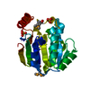 3l4eS S: Starting model for refinement |
|---|---|
| Similar structure data |


-Links
 PDBj
PDBj

- Assembly
| Deposited unit | 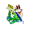
| ||||||||
|---|---|---|---|---|---|---|---|---|---|
| 1 |
| ||||||||
| Unit cell |
| ||||||||
| Components on special symmetry positions |
|
-Components
| #1: Protein | Mass: 28645.309 Da / Num. of mol.: 1 Source method: isolated from a genetically manipulated source Source: (gene. exp.) actinobacterium SCGC AAA027-L06 (bacteria)Gene: A27L6_002500000020 / Production host: |
|---|---|
| #2: Chemical | ChemComp-SO4 /   Mass: 96.063 Da / Num. of mol.: 1 / Source method: obtained synthetically / Formula: SO4 Mass: 96.063 Da / Num. of mol.: 1 / Source method: obtained synthetically / Formula: SO4 |
| #3: Water | ChemComp-HOH /  Mass: 18.015 Da / Num. of mol.: 163 / Source method: isolated from a natural source / Formula: H2O Mass: 18.015 Da / Num. of mol.: 163 / Source method: isolated from a natural source / Formula: H2O |
-Experimental details
-Experiment
| Experiment | Method: X-RAY DIFFRACTION / Number of used crystals: 1 |
|---|
- Sample preparation
| Crystal | Density Matthews: 2.28 Å3/Da / Density % sol: 46.04 % / Description: Long rods growing in clusters. |
|---|---|
| Crystal grow | Temperature: 293 K / Method: vapor diffusion, hanging drop / pH: 7.5 Details: Crystals of enzyme grew after mixing 2 microliters 16 mg/ml protein in buffer (30 mM maleic acid [pH 6.7], 200 mM Na2SO4) with an equal volume of condition 7 (100 mM HEPES-NaOH [pH 7.5], 2% ...Details: Crystals of enzyme grew after mixing 2 microliters 16 mg/ml protein in buffer (30 mM maleic acid [pH 6.7], 200 mM Na2SO4) with an equal volume of condition 7 (100 mM HEPES-NaOH [pH 7.5], 2% v/v PEG 400, 2 M (NH4)2SO4) from the TOP96 screen (Anatrace) over 500 microliters of the screen solution. 0.5 microliters IZIT crystal dye (Hampton) was added to the crystal-containing droplet after 4 weeks |
-Data collection
| Diffraction | Mean temperature: 100 K / Serial crystal experiment: N |
|---|---|
| Diffraction source | Source: SYNCHROTRON / Site: APS / Beamline: 21-ID-F / Wavelength: 0.97872 Å |
| Detector | Type: RAYONIX MX-300 / Detector: CCD / Date: Dec 8, 2018 |
| Radiation | Protocol: SINGLE WAVELENGTH / Monochromatic (M) / Laue (L): M / Scattering type: x-ray |
| Radiation wavelength | Wavelength: 0.97872 Å / Relative weight: 1 |
| Reflection | Resolution: 1.94→26.5 Å / Num. obs: 18380 / % possible obs: 91.6 % / Redundancy: 6.8 % / Biso Wilson estimate: 21.3 Å2 / CC1/2: 0.999 / Rpim(I) all: 0.034 / Rrim(I) all: 0.088 / Net I/σ(I): 26.4 |
| Reflection shell | Resolution: 1.94→1.97 Å / Redundancy: 6.8 % / Mean I/σ(I) obs: 3.14 / Num. unique obs: 953 / CC1/2: 0.915 / Rpim(I) all: 0.303 / Rrim(I) all: 0.805 / Χ2: 0.793 / % possible all: 98.8 |
- Processing
| Software |
| ||||||||||||||||||||||||||||||||||||||||||||||||||||||||
|---|---|---|---|---|---|---|---|---|---|---|---|---|---|---|---|---|---|---|---|---|---|---|---|---|---|---|---|---|---|---|---|---|---|---|---|---|---|---|---|---|---|---|---|---|---|---|---|---|---|---|---|---|---|---|---|---|---|
| Refinement | Method to determine structure: MOLECULAR REPLACEMENT Starting model: 3l4e Resolution: 1.94→26.409 Å / SU ML: 0.15 / Cross valid method: FREE R-VALUE / σ(F): 1.34 / Phase error: 20.68
| ||||||||||||||||||||||||||||||||||||||||||||||||||||||||
| Solvent computation | Shrinkage radii: 0.9 Å / VDW probe radii: 1.11 Å | ||||||||||||||||||||||||||||||||||||||||||||||||||||||||
| Displacement parameters | Biso mean: 27 Å2 | ||||||||||||||||||||||||||||||||||||||||||||||||||||||||
| Refinement step | Cycle: LAST / Resolution: 1.94→26.409 Å
| ||||||||||||||||||||||||||||||||||||||||||||||||||||||||
| Refine LS restraints |
| ||||||||||||||||||||||||||||||||||||||||||||||||||||||||
| LS refinement shell |
|
