 Movie
Movie Controller

[English] 日本語
 Yorodumi
- PDB-6nn4: The structure of human liver pyruvate kinase, hLPYK-D499N, in com... -
+ Open data

- Basic information
| Entry | Database: PDB / ID: 6nn4 | ||||||||||||||||||
|---|---|---|---|---|---|---|---|---|---|---|---|---|---|---|---|---|---|---|---|
| Title | The structure of human liver pyruvate kinase, hLPYK-D499N, in complex with Fru-1,6-BP | ||||||||||||||||||
 Components Components | Pyruvate kinase PKLR | ||||||||||||||||||
 Keywords Keywords | TRANSFERASE / pyruvate kinase / allosteric / glycolysis | ||||||||||||||||||
| Function / homology |  Function and homology information Function and homology informationpyruvate kinase complex / pyruvate biosynthetic process / SARS-CoV-1-host interactions / ChREBP activates metabolic gene expression / pyruvate kinase / pyruvate kinase activity / Pyruvate metabolism / monosaccharide binding / Glycolysis / response to metal ion ...pyruvate kinase complex / pyruvate biosynthetic process / SARS-CoV-1-host interactions / ChREBP activates metabolic gene expression / pyruvate kinase / pyruvate kinase activity / Pyruvate metabolism / monosaccharide binding / Glycolysis / response to metal ion / response to ATP / Regulation of gene expression in beta cells / potassium ion binding / response to cAMP / response to glucose / cellular response to epinephrine stimulus / response to nutrient / glycolytic process / cellular response to insulin stimulus / kinase activity / response to hypoxia / magnesium ion binding / extracellular exosome / ATP binding / cytosol / cytoplasm Similarity search - Function | ||||||||||||||||||
| Biological species |  Homo sapiens (human) Homo sapiens (human) | ||||||||||||||||||
| Method |  X-RAY DIFFRACTION / SYNCHROTRON / MOLECULAR REPLACEMENT / Resolution: 2.15 Å X-RAY DIFFRACTION / SYNCHROTRON / MOLECULAR REPLACEMENT / Resolution: 2.15 Å | ||||||||||||||||||
 Authors Authors | McFarlane, J.S. / Ronnebaum, T.A. / Meneely, K.M. / Fenton, A.W. / Lamb, A.L. | ||||||||||||||||||
| Funding support |  United States, 5items United States, 5items
| ||||||||||||||||||
 Citation Citation | Journal: Acta Crystallogr.,Sect.F / Year: 2019 Title: Changes in the allosteric site of human liver pyruvate kinase upon activator binding include the breakage of an intersubunit cation-pi bond. Authors: McFarlane, J.S. / Ronnebaum, T.A. / Meneely, K.M. / Chilton, A. / Fenton, A.W. / Lamb, A.L. | ||||||||||||||||||
| History |
|
- Structure visualization
| Structure viewer | Molecule: MolmilJmol/JSmol |
|---|
- Downloads & links
-Download
| PDBx/mmCIF format | 6nn4.cif.gz | 616.6 KB | Display | PDBx/mmCIF format |
|---|---|---|---|---|
| PDB format | pdb6nn4.ent.gz | 511.8 KB | Display | PDB format |
| PDBx/mmJSON format | 6nn4.json.gz | Tree view | PDBx/mmJSON format | |
| Others |  Other downloads Other downloads |
-Validation report
| Arichive directory | https://data.pdbj.org/pub/pdb/validation_reports/nn/6nn4ftp://data.pdbj.org/pub/pdb/validation_reports/nn/6nn4 | HTTPS FTP |
|---|
-Related structure data
| Related structure data |  6nn5C  6nn7C  6nn8C  4ip7S S: Starting model for refinement C: citing same article ( |
|---|---|
| Similar structure data |


-Links
 PDBj
PDBj


- Assembly
| Deposited unit | 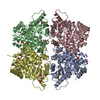
| ||||||||||||
|---|---|---|---|---|---|---|---|---|---|---|---|---|---|
| 1 |
| ||||||||||||
| Unit cell |
| ||||||||||||
| Components on special symmetry positions |
|
-Components
| #1: Protein | Mass: 58563.176 Da / Num. of mol.: 4 / Fragment: UNP residues 34-574 / Mutation: D499N Source method: isolated from a genetically manipulated source Source: (gene. exp.) Homo sapiens (human) / Gene: PKLR, PK1, PKL / Production host:  #2: Sugar | ChemComp-FBP /   Type: D-saccharide, beta linking / Mass: 340.116 Da / Num. of mol.: 4 Type: D-saccharide, beta linking / Mass: 340.116 Da / Num. of mol.: 4Source method: isolated from a genetically manipulated source Formula: C6H14O12P2 / Feature type: SUBJECT OF INVESTIGATION #3: Chemical | ChemComp-EDO /   Mass: 62.068 Da / Num. of mol.: 7 / Source method: obtained synthetically / Formula: C2H6O2 Mass: 62.068 Da / Num. of mol.: 7 / Source method: obtained synthetically / Formula: C2H6O2#4: Chemical | ChemComp-PEP /   Mass: 168.042 Da / Num. of mol.: 4 / Source method: obtained synthetically / Formula: C3H5O6P Mass: 168.042 Da / Num. of mol.: 4 / Source method: obtained synthetically / Formula: C3H5O6P#5: Water | ChemComp-HOH / |  Mass: 18.015 Da / Num. of mol.: 353 / Source method: isolated from a natural source / Formula: H2O Mass: 18.015 Da / Num. of mol.: 353 / Source method: isolated from a natural source / Formula: H2O |
|---|
-Experimental details
-Experiment
| Experiment | Method: X-RAY DIFFRACTION / Number of used crystals: 1 |
|---|
- Sample preparation
| Crystal | Density Matthews: 2.96 Å3/Da / Density % sol: 58.42 % Description: Crystals grew as rectangular prisms within two days and reached full size within two weeks. |
|---|---|
| Crystal grow | Temperature: 298 K / Method: vapor diffusion, hanging drop / Details: 0.2 M ammonium citrate dibasic, 20% PEG3350 |
-Data collection
| Diffraction | Mean temperature: 100 K / Serial crystal experiment: N |
|---|---|
| Diffraction source | Source: SYNCHROTRON / Site: SSRL / Beamline: BL12-2 / Wavelength: 0.97946 Å |
| Detector | Type: DECTRIS PILATUS3 6M / Detector: PIXEL / Date: Jan 28, 2017 |
| Radiation | Monochromator: double crystal Si(111) / Protocol: SINGLE WAVELENGTH / Monochromatic (M) / Laue (L): M / Scattering type: x-ray |
| Radiation wavelength | Wavelength: 0.97946 Å / Relative weight: 1 |
| Reflection | Resolution: 2.15→39.44 Å / Num. obs: 149968 / % possible obs: 99 % / Redundancy: 4.2 % / Net I/σ(I): 10.4 |
| Reflection shell | Resolution: 2.15→2.19 Å |
- Processing
| Software |
| |||||||||||||||||||||||||||||||||||||||||||||||||||||||||||||||||||||||||||||||||||||||||||||||||||||||||
|---|---|---|---|---|---|---|---|---|---|---|---|---|---|---|---|---|---|---|---|---|---|---|---|---|---|---|---|---|---|---|---|---|---|---|---|---|---|---|---|---|---|---|---|---|---|---|---|---|---|---|---|---|---|---|---|---|---|---|---|---|---|---|---|---|---|---|---|---|---|---|---|---|---|---|---|---|---|---|---|---|---|---|---|---|---|---|---|---|---|---|---|---|---|---|---|---|---|---|---|---|---|---|---|---|---|---|
| Refinement | Method to determine structure: MOLECULAR REPLACEMENT Starting model: PDB entry 4IP7 Resolution: 2.15→39.437 Å / SU ML: 0.28 / Cross valid method: FREE R-VALUE / σ(F): 1.34 / Phase error: 35.78
| |||||||||||||||||||||||||||||||||||||||||||||||||||||||||||||||||||||||||||||||||||||||||||||||||||||||||
| Solvent computation | Shrinkage radii: 0.9 Å / VDW probe radii: 1.11 Å | |||||||||||||||||||||||||||||||||||||||||||||||||||||||||||||||||||||||||||||||||||||||||||||||||||||||||
| Refinement step | Cycle: LAST / Resolution: 2.15→39.437 Å
| |||||||||||||||||||||||||||||||||||||||||||||||||||||||||||||||||||||||||||||||||||||||||||||||||||||||||
| Refine LS restraints |
| |||||||||||||||||||||||||||||||||||||||||||||||||||||||||||||||||||||||||||||||||||||||||||||||||||||||||
| LS refinement shell |
|
