 Movie
Movie Controller


+ Open data

- Basic information
| Entry | Database: PDB / ID: 6nm5 | |||||||||
|---|---|---|---|---|---|---|---|---|---|---|
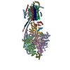
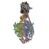
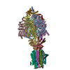
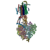
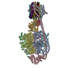
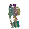
| Title | F-pilus/MS2 Maturation protein complex | |||||||||
 Components Components |
| |||||||||
 Keywords Keywords | PROTEIN BINDING / MS2 maturation protein / F-pilus / adsorption complex | |||||||||
| Function / homology |  Function and homology information Function and homology informationviral genome circularization / : / virion attachment to host cell pilus / membrane => GO:0016020 / virion component / RNA binding / extracellular region Similarity search - Function | |||||||||
| Biological species |   Enterobacteria phage MS2 (virus) Enterobacteria phage MS2 (virus) | |||||||||
| Method | ELECTRON MICROSCOPY / single particle reconstruction / cryo EM / Resolution: 6.2 Å | |||||||||
 Authors Authors | Meng, R. / Chang, J. / Zhang, J. | |||||||||
| Funding support |  United States, 2items United States, 2items
| |||||||||
 Citation Citation | Journal: Nat Commun / Year: 2019 Title: Structural basis for the adsorption of a single-stranded RNA bacteriophage. Authors: Ran Meng / Mengqiu Jiang / Zhicheng Cui / Jeng-Yih Chang / Kailu Yang / Joanita Jakana / Xinzhe Yu / Zhao Wang / Bo Hu / Junjie Zhang / Abstract: Single-stranded RNA bacteriophages (ssRNA phages) infect Gram-negative bacteria via a single maturation protein (Mat), which attaches to a retractile pilus of the host. Here we present structures of ...Single-stranded RNA bacteriophages (ssRNA phages) infect Gram-negative bacteria via a single maturation protein (Mat), which attaches to a retractile pilus of the host. Here we present structures of the ssRNA phage MS2 in complex with the Escherichia coli F-pilus, showing a network of hydrophobic and electrostatic interactions at the Mat-pilus interface. Moreover, binding of the pilus induces slight orientational variations of the Mat relative to the rest of the phage capsid, priming the Mat-connected genomic RNA (gRNA) for its release from the virions. The exposed tip of the attached Mat points opposite to the direction of the pilus retraction, which may facilitate the translocation of the gRNA from the capsid into the host cytosol. In addition, our structures determine the orientation of the assembled F-pilin subunits relative to the cell envelope, providing insights into the F-like type IV secretion systems. | |||||||||
| History |
|
- Structure visualization
| Movie |
Movie viewer |
|---|---|
| Structure viewer | Molecule: MolmilJmol/JSmol |
- Downloads & links
-Download
| PDBx/mmCIF format | 6nm5.cif.gz | 843.3 KB | Display | PDBx/mmCIF format |
|---|---|---|---|---|
| PDB format | pdb6nm5.ent.gz | Display | PDB format | |
| PDBx/mmJSON format | 6nm5.json.gz | Tree view | PDBx/mmJSON format | |
| Others |  Other downloads Other downloads |
-Validation report
| Arichive directory | https://data.pdbj.org/pub/pdb/validation_reports/nm/6nm5ftp://data.pdbj.org/pub/pdb/validation_reports/nm/6nm5 | HTTPS FTP |
|---|
-Related structure data
| Related structure data | 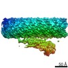 9397MC 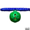 0338C 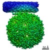 0448C  0450C 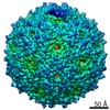 0451C 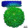 0453C 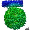 9399C C: citing same article ( M: map data used to model this data |
|---|---|
| Similar structure data |


-Links
 PDBj
PDBj- Assembly
| Deposited unit | 
|
|---|---|
| 1 |
|
-Components
| #1: Protein | Mass: 6831.216 Da / Num. of mol.: 75 / Source method: isolated from a natural source / Source: (natural) #2: Protein | | Mass: 44030.934 Da / Num. of mol.: 1 / Source method: isolated from a natural source / Source: (natural) Enterobacteria phage MS2 (virus) / References: UniProt: P03610#3: Chemical | ChemComp-KSV / (   Mass: 200.127 Da / Num. of mol.: 70 / Source method: obtained synthetically / Formula: C5H13O6P Mass: 200.127 Da / Num. of mol.: 70 / Source method: obtained synthetically / Formula: C5H13O6P |
|---|
-Experimental details
-Experiment
| Experiment | Method: ELECTRON MICROSCOPY |
|---|---|
| EM experiment | Aggregation state: PARTICLE / 3D reconstruction method: single particle reconstruction |
- Sample preparation
| Component |
| ||||||||||||||||||||||||||||
|---|---|---|---|---|---|---|---|---|---|---|---|---|---|---|---|---|---|---|---|---|---|---|---|---|---|---|---|---|---|
| Molecular weight | Experimental value: NO | ||||||||||||||||||||||||||||
| Source (natural) |
| ||||||||||||||||||||||||||||
| Buffer solution | pH: 8 | ||||||||||||||||||||||||||||
| Specimen | Embedding applied: NO / Shadowing applied: NO / Staining applied: NO / Vitrification applied: YES | ||||||||||||||||||||||||||||
| Specimen support | Grid material: COPPER / Grid mesh size: 400 divisions/in. / Grid type: C-flat-1.2/1.3 | ||||||||||||||||||||||||||||
| Vitrification | Cryogen name: ETHANE |
- Electron microscopy imaging
| Microscopy | Model: JEOL 3200FSC |
|---|---|
| Electron gun | Electron source:  FIELD EMISSION GUN / Accelerating voltage: 300 kV / Illumination mode: FLOOD BEAM FIELD EMISSION GUN / Accelerating voltage: 300 kV / Illumination mode: FLOOD BEAM |
| Electron lens | Mode: BRIGHT FIELD |
| Image recording | Average exposure time: 8 sec. / Electron dose: 37 e/Å2 / Film or detector model: GATAN K2 SUMMIT (4k x 4k) |
| EM imaging optics | Energyfilter slit width: 30 eV |
- Processing
| EM software |
| ||||||||||||||||||||||||
|---|---|---|---|---|---|---|---|---|---|---|---|---|---|---|---|---|---|---|---|---|---|---|---|---|---|
| CTF correction | Type: PHASE FLIPPING AND AMPLITUDE CORRECTION | ||||||||||||||||||||||||
| Symmetry | Point symmetry: C1 (asymmetric) | ||||||||||||||||||||||||
| 3D reconstruction | Resolution: 6.2 Å / Resolution method: FSC 0.143 CUT-OFF / Num. of particles: 168989 / Symmetry type: POINT |
