 Movie
Movie Controller


+ Open data

- Basic information
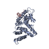
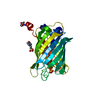
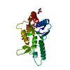
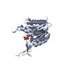
| Entry | Database: PDB / ID: 6nko | |||||||||
|---|---|---|---|---|---|---|---|---|---|---|
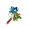
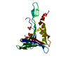
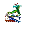
| Title | Crystal structure of ForH | |||||||||
 Components Components | ForH | |||||||||
 Keywords Keywords | UNKNOWN FUNCTION / Apo structure / Formycin A | |||||||||
| Function / homology |  Function and homology information Function and homology informationphosphoribosylaminoimidazolecarboxamide formyltransferase activity / IMP cyclohydrolase activity / purine nucleotide biosynthetic process Similarity search - Function | |||||||||
| Biological species |  Streptomyces kaniharaensis (bacteria) Streptomyces kaniharaensis (bacteria) | |||||||||
| Method |  X-RAY DIFFRACTION / SYNCHROTRON / MOLECULAR REPLACEMENT / Resolution: 2.403 Å X-RAY DIFFRACTION / SYNCHROTRON / MOLECULAR REPLACEMENT / Resolution: 2.403 Å | |||||||||
 Authors Authors | Zheng, J. / Irani, S. / Zhang, Y. | |||||||||
| Funding support |  United States, 2items United States, 2items
| |||||||||
 Citation Citation | Journal: J. Am. Chem. Soc. / Year: 2019 Title: Identification of the Formycin A Biosynthetic Gene Cluster from Streptomyces kaniharaensis Illustrates the Interplay between Biological Pyrazolopyrimidine Formation and de Novo Purine Biosynthesis. Authors: Wang, S.A. / Ko, Y. / Zeng, J. / Geng, Y. / Ren, D. / Ogasawara, Y. / Irani, S. / Zhang, Y. / Liu, H.W. | |||||||||
| History |
|
- Structure visualization
| Structure viewer | Molecule: MolmilJmol/JSmol |
|---|
- Downloads & links
-Download
| PDBx/mmCIF format | 6nko.cif.gz | 157.4 KB | Display | PDBx/mmCIF format |
|---|---|---|---|---|
| PDB format | pdb6nko.ent.gz | 123.9 KB | Display | PDB format |
| PDBx/mmJSON format | 6nko.json.gz | Tree view | PDBx/mmJSON format | |
| Others |  Other downloads Other downloads |
-Validation report
| Summary document | 6nko_validation.pdf.gz | 450.1 KB | Display | wwPDB validaton report |
|---|---|---|---|---|
| Full document | 6nko_full_validation.pdf.gz | 458.9 KB | Display | |
| Data in XML | 6nko_validation.xml.gz | 28.8 KB | Display | |
| Data in CIF | 6nko_validation.cif.gz | 39.4 KB | Display | |
| Arichive directory | https://data.pdbj.org/pub/pdb/validation_reports/nk/6nkoftp://data.pdbj.org/pub/pdb/validation_reports/nk/6nko | HTTPS FTP |
-Related structure data
| Related structure data |  1m9nS S: Starting model for refinement |
|---|---|
| Similar structure data |


-Links
 PDBj
PDBj
- Assembly
| Deposited unit | 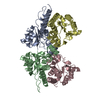
| ||||||||
|---|---|---|---|---|---|---|---|---|---|
| 1 | 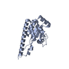
| ||||||||
| 2 | 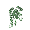
| ||||||||
| 3 | 
| ||||||||
| 4 | 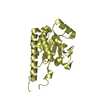
| ||||||||
| 5 | 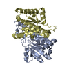
| ||||||||
| 6 | 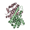
| ||||||||
| Unit cell |
|
-Components
| #1: Protein | Mass: 23192.391 Da / Num. of mol.: 4 Source method: isolated from a genetically manipulated source Source: (gene. exp.) Streptomyces kaniharaensis (bacteria) / Production host: #2: Water | ChemComp-HOH / |  Mass: 18.015 Da / Num. of mol.: 126 / Source method: isolated from a natural source / Formula: H2O Mass: 18.015 Da / Num. of mol.: 126 / Source method: isolated from a natural source / Formula: H2O |
|---|
-Experimental details
-Experiment
| Experiment | Method: X-RAY DIFFRACTION / Number of used crystals: 1 |
|---|
- Sample preparation
| Crystal | Density Matthews: 2.82 Å3/Da / Density % sol: 56.35 % |
|---|---|
| Crystal grow | Temperature: 295 K / Method: vapor diffusion, sitting drop / Details: 2 M ammonium sulfate, 0.1 M Bis-Tris, pH 5.5 |
-Data collection
| Diffraction | Mean temperature: 100 K / Serial crystal experiment: N |
|---|---|
| Diffraction source | Source: SYNCHROTRON / Site: APS / Beamline: 23-ID-B / Wavelength: 1.0332 Å |
| Detector | Type: DECTRIS EIGER X 16M / Detector: PIXEL / Date: May 22, 2016 |
| Radiation | Monochromator: Double crystal cryo-cooled Si(111) / Protocol: SINGLE WAVELENGTH / Monochromatic (M) / Laue (L): M / Scattering type: x-ray |
| Radiation wavelength | Wavelength: 1.0332 Å / Relative weight: 1 |
| Reflection | Resolution: 2.4→50 Å / Num. obs: 41344 / % possible obs: 100 % / Redundancy: 6.5 % / Net I/σ(I): 15.14 |
| Reflection shell | Resolution: 2.4→2.44 Å / Num. unique obs: 4551 |
- Processing
| Software |
| ||||||||||||||||||||||||||||||||||||||||||||||||||||||||||||||||||||||||||||||||||||||||||||||||||
|---|---|---|---|---|---|---|---|---|---|---|---|---|---|---|---|---|---|---|---|---|---|---|---|---|---|---|---|---|---|---|---|---|---|---|---|---|---|---|---|---|---|---|---|---|---|---|---|---|---|---|---|---|---|---|---|---|---|---|---|---|---|---|---|---|---|---|---|---|---|---|---|---|---|---|---|---|---|---|---|---|---|---|---|---|---|---|---|---|---|---|---|---|---|---|---|---|---|---|---|
| Refinement | Method to determine structure: MOLECULAR REPLACEMENT Starting model: PDB entry 1M9N Resolution: 2.403→48.93 Å / SU ML: 0.34 / Cross valid method: FREE R-VALUE / σ(F): 1.36 / Phase error: 27.65
| ||||||||||||||||||||||||||||||||||||||||||||||||||||||||||||||||||||||||||||||||||||||||||||||||||
| Solvent computation | Shrinkage radii: 0.9 Å / VDW probe radii: 1.11 Å | ||||||||||||||||||||||||||||||||||||||||||||||||||||||||||||||||||||||||||||||||||||||||||||||||||
| Refinement step | Cycle: LAST / Resolution: 2.403→48.93 Å
| ||||||||||||||||||||||||||||||||||||||||||||||||||||||||||||||||||||||||||||||||||||||||||||||||||
| Refine LS restraints |
| ||||||||||||||||||||||||||||||||||||||||||||||||||||||||||||||||||||||||||||||||||||||||||||||||||
| LS refinement shell |
|
