 Movie
Movie Controller

[English] 日本語
 Yorodumi
- PDB-6nf3: Structure of the Monoclinic-3 (Monocln-3) Crystal Form of Human A... -
+ Open data

- Basic information
| Entry | Database: PDB / ID: 6nf3 | ||||||
|---|---|---|---|---|---|---|---|
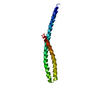
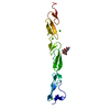
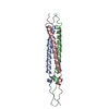
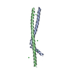
| Title | Structure of the Monoclinic-3 (Monocln-3) Crystal Form of Human Apolipoprotein C1 | ||||||
 Components Components | Apolipoprotein C-I | ||||||
 Keywords Keywords | LIPID BINDING PROTEIN / lipoprotein particles / cholesterol / blood / vascular | ||||||
| Function / homology |  Function and homology information Function and homology informationnegative regulation of phosphatidylcholine catabolic process / negative regulation of cholesterol transport / lipase inhibitor activity / negative regulation of very-low-density lipoprotein particle clearance / phospholipase inhibitor activity / VLDL assembly / plasma lipoprotein particle remodeling / regulation of cholesterol transport / very-low-density lipoprotein particle assembly / VLDL clearance ...negative regulation of phosphatidylcholine catabolic process / negative regulation of cholesterol transport / lipase inhibitor activity / negative regulation of very-low-density lipoprotein particle clearance / phospholipase inhibitor activity / VLDL assembly / plasma lipoprotein particle remodeling / regulation of cholesterol transport / very-low-density lipoprotein particle assembly / VLDL clearance / negative regulation of lipid metabolic process / negative regulation of triglyceride catabolic process / chylomicron remnant clearance / phosphatidylcholine-sterol O-acyltransferase activator activity / very-low-density lipoprotein particle clearance / phospholipid efflux / chylomicron / negative regulation of fatty acid biosynthetic process / high-density lipoprotein particle remodeling / lipoprotein metabolic process / phosphatidylcholine binding / high-density lipoprotein particle / very-low-density lipoprotein particle / triglyceride metabolic process / cholesterol efflux / triglyceride homeostasis / negative regulation of lipid catabolic process / cholesterol metabolic process / NR1H3 & NR1H2 regulate gene expression linked to cholesterol transport and efflux / fatty acid binding / lipid metabolic process / negative regulation of receptor-mediated endocytosis / endoplasmic reticulum / extracellular region Similarity search - Function | ||||||
| Biological species |  Homo sapiens (human) Homo sapiens (human) | ||||||
| Method |  X-RAY DIFFRACTION / MOLECULAR REPLACEMENT / Resolution: 2.33 Å X-RAY DIFFRACTION / MOLECULAR REPLACEMENT / Resolution: 2.33 Å | ||||||
 Authors Authors | McPherson, A. / Larson, S.B. | ||||||
 Citation Citation | Journal: J. Lipid Res. / Year: 2019 Title: The structure of human apolipoprotein C-1 in four different crystal forms. Authors: McPherson, A. / Larson, S.B. | ||||||
| History |
|
- Structure visualization
| Structure viewer | Molecule: MolmilJmol/JSmol |
|---|
- Downloads & links
-Download
| PDBx/mmCIF format | 6nf3.cif.gz | 36.6 KB | Display | PDBx/mmCIF format |
|---|---|---|---|---|
| PDB format | pdb6nf3.ent.gz | 24.1 KB | Display | PDB format |
| PDBx/mmJSON format | 6nf3.json.gz | Tree view | PDBx/mmJSON format | |
| Others |  Other downloads Other downloads |
-Validation report
| Arichive directory | https://data.pdbj.org/pub/pdb/validation_reports/nf/6nf3ftp://data.pdbj.org/pub/pdb/validation_reports/nf/6nf3 | HTTPS FTP |
|---|
-Related structure data
| Related structure data |  6dvuC  6dxrC  6dz6C  1ropS S: Starting model for refinement C: citing same article ( |
|---|---|
| Similar structure data |

-Links
 PDBj
PDBj


- Assembly
| Deposited unit | 
| ||||||||
|---|---|---|---|---|---|---|---|---|---|
| 1 | 
| ||||||||
| Unit cell |
|
-Components
| #1: Protein | Mass: 9344.909 Da / Num. of mol.: 2 / Source method: isolated from a natural source / Source: (natural) Homo sapiens (human) / References: UniProt: P02654#2: Water | ChemComp-HOH / |  Mass: 18.015 Da / Num. of mol.: 108 / Source method: isolated from a natural source / Formula: H2O Mass: 18.015 Da / Num. of mol.: 108 / Source method: isolated from a natural source / Formula: H2O |
|---|
-Experimental details
-Experiment
| Experiment | Method: X-RAY DIFFRACTION / Number of used crystals: 1 |
|---|
- Sample preparation
| Crystal | Density Matthews: 1.74 Å3/Da / Density % sol: 29.51 % / Description: thin laths |
|---|---|
| Crystal grow | Temperature: 298 K / Method: vapor diffusion / pH: 6.5 Details: The crystals were grown by sitting drop vapor diffusion in Cryschem plates using 0.6 ml reservoirs of 16% to 18% 2-methyl-2,4-pentanediol (MPD) containing o.1 M sodium acetate and 0.25% ...Details: The crystals were grown by sitting drop vapor diffusion in Cryschem plates using 0.6 ml reservoirs of 16% to 18% 2-methyl-2,4-pentanediol (MPD) containing o.1 M sodium acetate and 0.25% octyl-beta-s-1-thioglucopyanoside. The drops were equal volumes, generally 6 ul each, of the reservoir and an 8 mg/ml solution of protein dissolved in .02 M ammonium bicarbonate. PH range: 6.0 7.0 |
-Data collection
| Diffraction | Mean temperature: 298 K | |||||||||||||||
|---|---|---|---|---|---|---|---|---|---|---|---|---|---|---|---|---|
| Diffraction source | Source: ROTATING ANODE / Type: RIGAKU FR-D / Wavelength: 1.54 Å | |||||||||||||||
| Detector | Type: SDMS / Detector: AREA DETECTOR / Date: Jul 15, 1992 | |||||||||||||||
| Radiation | Monochromator: Supper / Protocol: SINGLE WAVELENGTH / Monochromatic (M) / Laue (L): M / Scattering type: x-ray | |||||||||||||||
| Radiation wavelength | Wavelength: 1.54 Å / Relative weight: 1 | |||||||||||||||
| Reflection twin |
| |||||||||||||||
| Reflection | Resolution: 2.2→30 Å / Num. obs: 5846 / % possible obs: 70 % / Redundancy: 2.6 % / CC1/2: 0.987 / Rmerge(I) obs: 0.131 / Rpim(I) all: 0.071 / Rrim(I) all: 0.15 / Rsym value: 0.131 / Net I/av σ(I): 4.6 / Net I/σ(I): 4.6 | |||||||||||||||
| Reflection shell | Resolution: 2.4→2.5 Å / Redundancy: 1.2 % / Rmerge(I) obs: 0.211 / Mean I/σ(I) obs: 1.7 / Num. unique obs: 263 / CC1/2: 0.933 / Rpim(I) all: 0.206 / Rrim(I) all: 0.295 / Rsym value: 0.211 / % possible all: 46.2 |
- Processing
| Software |
| ||||||||||||||||||||||||||||||||||||||||||||||||||||||||||||||||||||||||||||||||||||||||||||||||||||||||||||||||||||||||||||||||||||||||||||||||||||||||||||||||||||||||||||||||||||||
|---|---|---|---|---|---|---|---|---|---|---|---|---|---|---|---|---|---|---|---|---|---|---|---|---|---|---|---|---|---|---|---|---|---|---|---|---|---|---|---|---|---|---|---|---|---|---|---|---|---|---|---|---|---|---|---|---|---|---|---|---|---|---|---|---|---|---|---|---|---|---|---|---|---|---|---|---|---|---|---|---|---|---|---|---|---|---|---|---|---|---|---|---|---|---|---|---|---|---|---|---|---|---|---|---|---|---|---|---|---|---|---|---|---|---|---|---|---|---|---|---|---|---|---|---|---|---|---|---|---|---|---|---|---|---|---|---|---|---|---|---|---|---|---|---|---|---|---|---|---|---|---|---|---|---|---|---|---|---|---|---|---|---|---|---|---|---|---|---|---|---|---|---|---|---|---|---|---|---|---|---|---|---|---|
| Refinement | Method to determine structure: MOLECULAR REPLACEMENT Starting model: 1ROP Resolution: 2.33→18.97 Å / Cor.coef. Fo:Fc: 0.832 / Cor.coef. Fo:Fc free: 0.618 / SU B: 13.65 / SU ML: 0.317 / Cross valid method: THROUGHOUT / ESU R: 0.183 / ESU R Free: 0.083 / Stereochemistry target values: MAXIMUM LIKELIHOOD / Details: HYDROGENS HAVE BEEN ADDED IN THE RIDING POSITIONS
| ||||||||||||||||||||||||||||||||||||||||||||||||||||||||||||||||||||||||||||||||||||||||||||||||||||||||||||||||||||||||||||||||||||||||||||||||||||||||||||||||||||||||||||||||||||||
| Solvent computation | Ion probe radii: 0.8 Å / Shrinkage radii: 0.8 Å / VDW probe radii: 1.2 Å / Solvent model: BABINET MODEL WITH MASK | ||||||||||||||||||||||||||||||||||||||||||||||||||||||||||||||||||||||||||||||||||||||||||||||||||||||||||||||||||||||||||||||||||||||||||||||||||||||||||||||||||||||||||||||||||||||
| Displacement parameters | Biso mean: 21.243 Å2
| ||||||||||||||||||||||||||||||||||||||||||||||||||||||||||||||||||||||||||||||||||||||||||||||||||||||||||||||||||||||||||||||||||||||||||||||||||||||||||||||||||||||||||||||||||||||
| Refinement step | Cycle: 1 / Resolution: 2.33→18.97 Å
| ||||||||||||||||||||||||||||||||||||||||||||||||||||||||||||||||||||||||||||||||||||||||||||||||||||||||||||||||||||||||||||||||||||||||||||||||||||||||||||||||||||||||||||||||||||||
| Refine LS restraints |
|