 Movie
Movie Controller

[English] 日本語
 Yorodumi
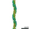
- PDB-6nef: Outer Membrane Cytochrome S Filament from Geobacter Sulfurreducens -
+ Open data

- Basic information
| Entry | Database: PDB / ID: 6nef | |||||||||
|---|---|---|---|---|---|---|---|---|---|---|
| Title | Outer Membrane Cytochrome S Filament from Geobacter Sulfurreducens | |||||||||
 Components Components | C-type cytochrome OmcS | |||||||||
 Keywords Keywords | PROTEIN FIBRIL / Conductive / filament / nanowire / SIX-HEME MULTIHEME C-TYPE CYTOCHROME | |||||||||
| Function / homology | : / Multiheme cytochrome superfamily / anaerobic respiration / cell outer membrane / electron transfer activity / cell surface / metal ion binding / HEME C / C-type cytochrome OmcS Function and homology information Function and homology information | |||||||||
| Biological species |  Geobacter sulfurreducens (bacteria) Geobacter sulfurreducens (bacteria) | |||||||||
| Method | ELECTRON MICROSCOPY / helical reconstruction / cryo EM / Resolution: 3.4 Å | |||||||||
 Authors Authors | Filman, D.J. / Marino, S.F. / Ward, J.E. / Yang, L. / Mester, Z. / Bullitt, E. / Lovley, D.R. / Strauss, M. | |||||||||
| Funding support |  United States, 1items United States, 1items
| |||||||||
 Citation Citation | Journal: Commun Biol / Year: 2019 Title: Cryo-EM reveals the structural basis of long-range electron transport in a cytochrome-based bacterial nanowire. Authors: David J Filman / Stephen F Marino / Joy E Ward / Lu Yang / Zoltán Mester / Esther Bullitt / Derek R Lovley / Mike Strauss /   Abstract: Electrically conductive pili from species, termed bacterial nanowires, are intensely studied for their biological significance and potential in the development of new materials. Using cryo-electron ...Electrically conductive pili from species, termed bacterial nanowires, are intensely studied for their biological significance and potential in the development of new materials. Using cryo-electron microscopy, we have characterized nanowires from conductive pili preparations that are composed solely of head-to-tail stacked monomers of the six-heme C-type cytochrome OmcS. The unique fold of OmcS - closely wrapped around a continuous stack of hemes that can serve as an uninterrupted path for electron transport - generates a scaffold that supports the unbranched chain of hemes along the central axis of the filament. We present here, at 3.4 Å resolution, the structure of this cytochrome-based filament and discuss its possible role in long-range biological electron transport. | |||||||||
| History |
|
- Structure visualization
| Movie |
Movie viewer |
|---|---|
| Structure viewer | Molecule: MolmilJmol/JSmol |
- Downloads & links
-Download
| PDBx/mmCIF format | 6nef.cif.gz | 103 KB | Display | PDBx/mmCIF format |
|---|---|---|---|---|
| PDB format | pdb6nef.ent.gz | 75.1 KB | Display | PDB format |
| PDBx/mmJSON format | 6nef.json.gz | Tree view | PDBx/mmJSON format | |
| Others |  Other downloads Other downloads |
-Validation report
| Summary document | 6nef_validation.pdf.gz | 1.1 MB | Display | wwPDB validaton report |
|---|---|---|---|---|
| Full document | 6nef_full_validation.pdf.gz | 1.1 MB | Display | |
| Data in XML | 6nef_validation.xml.gz | 23.6 KB | Display | |
| Data in CIF | 6nef_validation.cif.gz | 32.2 KB | Display | |
| Arichive directory | https://data.pdbj.org/pub/pdb/validation_reports/ne/6nefftp://data.pdbj.org/pub/pdb/validation_reports/ne/6nef | HTTPS FTP |
-Related structure data
| Related structure data | 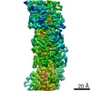 9357MC M: map data used to model this data C: citing same article ( |
|---|---|
| Similar structure data |
-Links
 PDBj
PDBj


- Assembly
| Deposited unit | 
|
|---|---|
| 1 | x 9
|
| 2 |
|
| Symmetry | Helical symmetry: (Circular symmetry: 1 / Dyad axis: no / N subunits divisor: 1 / Num. of operations: 9 / Rise per n subunits: 47.5 Å / Rotation per n subunits: 83.01 °) |
| Details | SOME OBSERVED FILAMENTS ARE HUNDREDS OF SUBUNITS LONG |
-Components
| #1: Protein | Mass: 42986.008 Da / Num. of mol.: 1 / Source method: isolated from a natural source Details: SIX-HEME MULTIHEME C-TYPE CYTOCHROME, PROTEIN FIBRIL Source: (natural) Geobacter sulfurreducens (strain ATCC 51573 / DSM 12127 / PCA) (bacteria)Strain: ATCC 51573 / DSM 12127 / PCA / References: UniProt: Q74A86 | ||||
|---|---|---|---|---|---|
| #2: Chemical | ChemComp-HEC /   Mass: 618.503 Da / Num. of mol.: 6 / Source method: obtained synthetically / Formula: C34H34FeN4O4 Mass: 618.503 Da / Num. of mol.: 6 / Source method: obtained synthetically / Formula: C34H34FeN4O4#3: Chemical | ChemComp-MG / |   Mass: 24.305 Da / Num. of mol.: 1 / Source method: obtained synthetically / Formula: Mg Mass: 24.305 Da / Num. of mol.: 1 / Source method: obtained synthetically / Formula: MgHas protein modification | Y | |
-Experimental details
-Experiment
| Experiment | Method: ELECTRON MICROSCOPY |
|---|---|
| EM experiment | Aggregation state: FILAMENT / 3D reconstruction method: helical reconstruction |
- Sample preparation
| Component | Name: Outer Membrane Cytochrome S filament as a bacterial nanowire Type: ORGANELLE OR CELLULAR COMPONENT / Entity ID: #1 / Source: NATURAL |
|---|---|
| Molecular weight | Experimental value: NO |
| Source (natural) | Organism: Geobacter sulfurreducens (bacteria) |
| Buffer solution | pH: 7 |
| Specimen | Embedding applied: NO / Shadowing applied: NO / Staining applied: NO / Vitrification applied: YES Details: Sample contained thick (4nm) and thin (3nm) filaments. This reconstruction is of the thick filaments. |
| Vitrification | Instrument: FEI VITROBOT MARK IV / Cryogen name: ETHANE / Humidity: 100 % / Chamber temperature: 277 K |
- Electron microscopy imaging
| Experimental equipment |  Model: Titan Krios / Image courtesy: FEI Company |
|---|---|
| Microscopy | Model: FEI TITAN KRIOS |
| Electron gun | Electron source:  FIELD EMISSION GUN / Accelerating voltage: 300 kV / Illumination mode: FLOOD BEAM FIELD EMISSION GUN / Accelerating voltage: 300 kV / Illumination mode: FLOOD BEAM |
| Electron lens | Mode: BRIGHT FIELD |
| Image recording | Electron dose: 45 e/Å2 / Detector mode: COUNTING / Film or detector model: GATAN K2 QUANTUM (4k x 4k) |
- Processing
| Software | Name: REFMAC / Version: 5.8.0238 / Classification: refinement | ||||||||||||||||||||||||||||||||||||||||||||||||||||||||||||||||||||||||||||||||||||||||||||||||||||||||||
|---|---|---|---|---|---|---|---|---|---|---|---|---|---|---|---|---|---|---|---|---|---|---|---|---|---|---|---|---|---|---|---|---|---|---|---|---|---|---|---|---|---|---|---|---|---|---|---|---|---|---|---|---|---|---|---|---|---|---|---|---|---|---|---|---|---|---|---|---|---|---|---|---|---|---|---|---|---|---|---|---|---|---|---|---|---|---|---|---|---|---|---|---|---|---|---|---|---|---|---|---|---|---|---|---|---|---|---|
| EM software |
| ||||||||||||||||||||||||||||||||||||||||||||||||||||||||||||||||||||||||||||||||||||||||||||||||||||||||||
| CTF correction | Type: PHASE FLIPPING AND AMPLITUDE CORRECTION | ||||||||||||||||||||||||||||||||||||||||||||||||||||||||||||||||||||||||||||||||||||||||||||||||||||||||||
| Helical symmerty | Angular rotation/subunit: 83.01 ° / Axial rise/subunit: 47.5 Å / Axial symmetry: C1 | ||||||||||||||||||||||||||||||||||||||||||||||||||||||||||||||||||||||||||||||||||||||||||||||||||||||||||
| 3D reconstruction | Resolution: 3.4 Å / Resolution method: FSC 0.143 CUT-OFF / Num. of particles: 462964 / Symmetry type: HELICAL | ||||||||||||||||||||||||||||||||||||||||||||||||||||||||||||||||||||||||||||||||||||||||||||||||||||||||||
| Atomic model building | Protocol: OTHER / Space: RECIPROCAL / Target criteria: maximum likelihood with phases Details: Atomic model was built de novo visually and repeatedly rebuilt visually using Coot and SPDBV. After each round of manual rebuilding, the parameters of the atomic model underwent ...Details: Atomic model was built de novo visually and repeatedly rebuilt visually using Coot and SPDBV. After each round of manual rebuilding, the parameters of the atomic model underwent stereochemically restrained reciprocal space refinement, using either Refmac5 or Phenix.autobuild or both, using as a reference the amplitudes and phases of the Fourier transform of a portion of the cryoEM reconstruction. For convenience, a single-subunit model was initially built to fit an approximation of the reference map in space group P4(3), which was subsequently replaced by a three-subunit model, using the authentic helical parameters of the map. one of the two axial ligands for HEM 505 is ne2 of his 41 from a neighboring helical-symmetry-related protein chain. | ||||||||||||||||||||||||||||||||||||||||||||||||||||||||||||||||||||||||||||||||||||||||||||||||||||||||||
| Refinement | Highest resolution: 3.4 Å Details: ONE OF THE TWO AXIAL LIGANDS FOR HEM 505 IS NE2 OF HIS 41 FROM A NEIGHBORING HELICAL-SYMMETRY-RELATED PROTEIN CHAIN | ||||||||||||||||||||||||||||||||||||||||||||||||||||||||||||||||||||||||||||||||||||||||||||||||||||||||||
| Refinement step | Cycle: 1 / Total: 9648 | ||||||||||||||||||||||||||||||||||||||||||||||||||||||||||||||||||||||||||||||||||||||||||||||||||||||||||
| Refine LS restraints |
|