 Movie
Movie Controller

[English] 日本語
 Yorodumi
- PDB-6nbw: Ternary Complex of Beta/Gamma-Actin with Profilin and AnCoA-NAA80 -
+ Open data

- Basic information
| Entry | Database: PDB / ID: 6nbw | ||||||
|---|---|---|---|---|---|---|---|
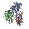
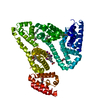
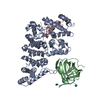
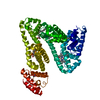
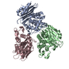
| Title | Ternary Complex of Beta/Gamma-Actin with Profilin and AnCoA-NAA80 | ||||||
 Components Components |
| ||||||
 Keywords Keywords | STRUCTURAL PROTEIN/TRANSFERASE / acetylation / NAA80 / CYTOSOLIC PROTEIN / STRUCTURAL PROTEIN-TRANSFERASE complex | ||||||
| Function / homology |  Function and homology information Function and homology informationN-terminal peptidyl-aspartic acid acetylation / N-terminal peptidyl-glutamic acid acetylation / actin modification / acetyl-CoA binding / regulation of actin polymerization or depolymerization / synapse maturation / protein-N-terminal amino-acid acetyltransferase activity / adenyl-nucleotide exchange factor activity / modification of postsynaptic actin cytoskeleton / positive regulation of norepinephrine uptake ...N-terminal peptidyl-aspartic acid acetylation / N-terminal peptidyl-glutamic acid acetylation / actin modification / acetyl-CoA binding / regulation of actin polymerization or depolymerization / synapse maturation / protein-N-terminal amino-acid acetyltransferase activity / adenyl-nucleotide exchange factor activity / modification of postsynaptic actin cytoskeleton / positive regulation of norepinephrine uptake / negative regulation of actin filament bundle assembly / positive regulation of actin filament bundle assembly / negative regulation of actin filament polymerization / bBAF complex / cellular response to cytochalasin B / regulation of actin filament polymerization / npBAF complex / N-acetyltransferase activity / nBAF complex / brahma complex / Signaling by ROBO receptors / regulation of transepithelial transport / Formation of annular gap junctions / morphogenesis of a polarized epithelium / Formation of the dystrophin-glycoprotein complex (DGC) / structural constituent of postsynaptic actin cytoskeleton / GBAF complex / Gap junction degradation / regulation of G0 to G1 transition / Folding of actin by CCT/TriC / Cell-extracellular matrix interactions / protein localization to adherens junction / dense body / positive regulation of ATP-dependent activity / postsynaptic actin cytoskeleton / Tat protein binding / Prefoldin mediated transfer of substrate to CCT/TriC / RSC-type complex / regulation of double-strand break repair / regulation of nucleotide-excision repair / proline-rich region binding / PCP/CE pathway / Adherens junctions interactions / RHOF GTPase cycle / adherens junction assembly / apical protein localization / positive regulation of ruffle assembly / Sensory processing of sound by outer hair cells of the cochlea / negative regulation of stress fiber assembly / Interaction between L1 and Ankyrins / tight junction / SWI/SNF complex / regulation of mitotic metaphase/anaphase transition / Sensory processing of sound by inner hair cells of the cochlea / positive regulation of T cell differentiation / protein acetylation / apical junction complex / regulation of norepinephrine uptake / positive regulation of double-strand break repair / transporter regulator activity / maintenance of blood-brain barrier / nitric-oxide synthase binding / cortical cytoskeleton / positive regulation of actin filament polymerization / establishment or maintenance of cell polarity / NuA4 histone acetyltransferase complex / positive regulation of stem cell population maintenance / Regulation of MITF-M-dependent genes involved in pigmentation / Recycling pathway of L1 / brush border / regulation of G1/S transition of mitotic cell cycle / positive regulation of epithelial cell migration / actin monomer binding / kinesin binding / EPH-ephrin mediated repulsion of cells / negative regulation of cell differentiation / RHO GTPases Activate WASPs and WAVEs / regulation of synaptic vesicle endocytosis / positive regulation of myoblast differentiation / RHO GTPases activate IQGAPs / regulation of protein localization to plasma membrane / positive regulation of double-strand break repair via homologous recombination / phosphatidylinositol-4,5-bisphosphate binding / phosphotyrosine residue binding / EPHB-mediated forward signaling / Transferases; Acyltransferases; Transferring groups other than aminoacyl groups / cytoskeleton organization / substantia nigra development / axonogenesis / calyx of Held / nitric-oxide synthase regulator activity / neural tube closure / Translocation of SLC2A4 (GLUT4) to the plasma membrane / adherens junction / FCGR3A-mediated phagocytosis / actin filament / Regulation of endogenous retroelements by Piwi-interacting RNAs (piRNAs) / positive regulation of cell differentiation / cell motility / RHO GTPases Activate Formins Similarity search - Function | ||||||
| Biological species |  Homo sapiens (human) Homo sapiens (human) | ||||||
| Method |  X-RAY DIFFRACTION / SYNCHROTRON / MOLECULAR REPLACEMENT / Resolution: 2.5 Å X-RAY DIFFRACTION / SYNCHROTRON / MOLECULAR REPLACEMENT / Resolution: 2.5 Å | ||||||
 Authors Authors | Rebowski, G. / Boczkowska, M. / Dominguez, R. | ||||||
| Funding support |  United States, 1items United States, 1items
| ||||||
 Citation Citation | Journal: Sci Adv / Year: 2020 Title: Mechanism of actin N-terminal acetylation. Authors: Rebowski, G. / Boczkowska, M. / Drazic, A. / Ree, R. / Goris, M. / Arnesen, T. / Dominguez, R. | ||||||
| History |
|
- Structure visualization
| Structure viewer | Molecule: MolmilJmol/JSmol |
|---|
- Downloads & links
-Download
| PDBx/mmCIF format | 6nbw.cif.gz | 273.7 KB | Display | PDBx/mmCIF format |
|---|---|---|---|---|
| PDB format | pdb6nbw.ent.gz | 218 KB | Display | PDB format |
| PDBx/mmJSON format | 6nbw.json.gz | Tree view | PDBx/mmJSON format | |
| Others |  Other downloads Other downloads |
-Validation report
| Summary document | 6nbw_validation.pdf.gz | 1.4 MB | Display | wwPDB validaton report |
|---|---|---|---|---|
| Full document | 6nbw_full_validation.pdf.gz | 1.4 MB | Display | |
| Data in XML | 6nbw_validation.xml.gz | 28.9 KB | Display | |
| Data in CIF | 6nbw_validation.cif.gz | 39.7 KB | Display | |
| Arichive directory | https://data.pdbj.org/pub/pdb/validation_reports/nb/6nbwftp://data.pdbj.org/pub/pdb/validation_reports/nb/6nbw | HTTPS FTP |
-Related structure data
| Related structure data |  6nasC  6nbeC  2pbdS S: Starting model for refinement C: citing same article ( |
|---|---|
| Similar structure data |


-Links
 PDBj
PDBj


- Assembly
| Deposited unit | 
| ||||||||
|---|---|---|---|---|---|---|---|---|---|
| 1 |
| ||||||||
| Unit cell |
|
-Components
-Protein , 3 types, 3 molecules ANP
| #1: Protein | Mass: 41664.484 Da / Num. of mol.: 1 / Source method: isolated from a natural source / Source: (natural) Homo sapiens (human) / References: UniProt: P60709 |
|---|---|
| #2: Protein | Mass: 25923.076 Da / Num. of mol.: 1 / Fragment: residues 56-286 Source method: isolated from a genetically manipulated source Source: (gene. exp.) Homo sapiens (human) / Gene: NAA80, FUS2, NAT6 / Production host:  References: UniProt: Q93015, Transferases; Acyltransferases; Transferring groups other than aminoacyl groups |
| #3: Protein | Mass: 15071.222 Da / Num. of mol.: 1 Source method: isolated from a genetically manipulated source Source: (gene. exp.) Homo sapiens (human) / Gene: PFN1 / Production host: |
-Non-polymers , 7 types, 211 molecules 


| #4: Chemical | ChemComp-CA /  Mass: 40.078 Da / Num. of mol.: 1 / Source method: isolated from a natural source / Formula: Ca Mass: 40.078 Da / Num. of mol.: 1 / Source method: isolated from a natural source / Formula: Ca | ||||||
|---|---|---|---|---|---|---|---|
| #5: Chemical | ChemComp-ATP /  Mass: 507.181 Da / Num. of mol.: 1 / Source method: obtained synthetically / Formula: C10H16N5O13P3 / Comment: ATP, energy-carrying molecule*YM Mass: 507.181 Da / Num. of mol.: 1 / Source method: obtained synthetically / Formula: C10H16N5O13P3 / Comment: ATP, energy-carrying molecule*YM | ||||||
| #6: Chemical | ChemComp-LAB /  Mass: 395.513 Da / Num. of mol.: 1 / Source method: obtained synthetically / Formula: C20H29NO5S Mass: 395.513 Da / Num. of mol.: 1 / Source method: obtained synthetically / Formula: C20H29NO5S | ||||||
| #7: Chemical |  Mass: 92.094 Da / Num. of mol.: 2 / Source method: obtained synthetically / Formula: C3H8O3 Mass: 92.094 Da / Num. of mol.: 2 / Source method: obtained synthetically / Formula: C3H8O3#8: Chemical | ChemComp-SOP / [( |  Mass: 823.597 Da / Num. of mol.: 1 / Source method: obtained synthetically / Formula: C24H40N7O17P3S Mass: 823.597 Da / Num. of mol.: 1 / Source method: obtained synthetically / Formula: C24H40N7O17P3S#9: Chemical | ChemComp-MES / |  Mass: 195.237 Da / Num. of mol.: 1 / Source method: obtained synthetically / Formula: C6H13NO4S / Comment: pH buffer*YM Mass: 195.237 Da / Num. of mol.: 1 / Source method: obtained synthetically / Formula: C6H13NO4S / Comment: pH buffer*YM#10: Water | ChemComp-HOH / | Mass: 18.015 Da / Num. of mol.: 204 / Source method: isolated from a natural source / Formula: H2O |
-Details
| Has ligand of interest | N |
|---|
-Experimental details
-Experiment
| Experiment | Method: X-RAY DIFFRACTION / Number of used crystals: 1 |
|---|
- Sample preparation
| Crystal | Density Matthews: 2.65 Å3/Da / Density % sol: 53.56 % |
|---|---|
| Crystal grow | Temperature: 290 K / Method: vapor diffusion, hanging drop / pH: 6.5 / Details: 15% PEG 3350, 50mM MES pH6.2, 50mM NH4NO3 |
-Data collection
| Diffraction | Mean temperature: 100 K / Serial crystal experiment: N |
|---|---|
| Diffraction source | Source: SYNCHROTRON / Site: CHESS / Beamline: F1 / Wavelength: 0.9775 Å |
| Detector | Type: DECTRIS PILATUS 6M / Detector: PIXEL / Date: Nov 27, 2017 |
| Radiation | Protocol: SINGLE WAVELENGTH / Monochromatic (M) / Laue (L): M / Scattering type: x-ray |
| Radiation wavelength | Wavelength: 0.9775 Å / Relative weight: 1 |
| Reflection | Resolution: 2.496→50 Å / Num. obs: 28132 / % possible obs: 100 % / Redundancy: 13.1 % / Biso Wilson estimate: 33 Å2 / Rmerge(I) obs: 0.116 / Net I/σ(I): 21 |
| Reflection shell | Resolution: 2.5→2.59 Å / Redundancy: 13.3 % / Rmerge(I) obs: 0.563 / Mean I/σ(I) obs: 4 / Num. unique obs: 2785 / % possible all: 100 |
- Processing
| Software |
| |||||||||||||||||||||||||||||||||||||||||||||||||||||||||||||||||||||||||||||
|---|---|---|---|---|---|---|---|---|---|---|---|---|---|---|---|---|---|---|---|---|---|---|---|---|---|---|---|---|---|---|---|---|---|---|---|---|---|---|---|---|---|---|---|---|---|---|---|---|---|---|---|---|---|---|---|---|---|---|---|---|---|---|---|---|---|---|---|---|---|---|---|---|---|---|---|---|---|---|
| Refinement | Method to determine structure: MOLECULAR REPLACEMENT Starting model: 2PBD Resolution: 2.5→26.1 Å / SU ML: 0.26 / Cross valid method: THROUGHOUT / σ(F): 1.36 / Phase error: 20.17 / Stereochemistry target values: ML
| |||||||||||||||||||||||||||||||||||||||||||||||||||||||||||||||||||||||||||||
| Solvent computation | Shrinkage radii: 0.9 Å / VDW probe radii: 1.11 Å / Solvent model: FLAT BULK SOLVENT MODEL | |||||||||||||||||||||||||||||||||||||||||||||||||||||||||||||||||||||||||||||
| Displacement parameters | Biso max: 138.16 Å2 / Biso mean: 43.413 Å2 / Biso min: 24.15 Å2 | |||||||||||||||||||||||||||||||||||||||||||||||||||||||||||||||||||||||||||||
| Refinement step | Cycle: final / Resolution: 2.5→26.1 Å
| |||||||||||||||||||||||||||||||||||||||||||||||||||||||||||||||||||||||||||||
| LS refinement shell | Refine-ID: X-RAY DIFFRACTION / Rfactor Rfree error: 0 / Total num. of bins used: 10
|
