de novo centriole assembly involved in multi-ciliated epithelial cell differentiation / procentriole / deuterosome / procentriole replication complex / positive regulation of centriole replication / cell competition in a multicellular organism / histone H2AT120 kinase activity / trophoblast giant cell differentiation / polo kinase / V(D)J recombination ...de novo centriole assembly involved in multi-ciliated epithelial cell differentiation / procentriole / deuterosome / procentriole replication complex / positive regulation of centriole replication / cell competition in a multicellular organism / histone H2AT120 kinase activity / trophoblast giant cell differentiation / polo kinase / V(D)J recombination / XY body / Cul4-RING E3 ubiquitin ligase complex / centriole replication / cleavage furrow / cilium assembly / ubiquitin-like ligase-substrate adaptor activity / Loss of Nlp from mitotic centrosomes / Loss of proteins required for interphase microtubule organization from the centrosome / Recruitment of mitotic centrosome proteins and complexes / Recruitment of NuMA to mitotic centrosomes / Anchoring of the basal body to the plasma membrane / post-translational protein modification / B cell differentiation / AURKA Activation by TPX2 / nuclear estrogen receptor binding / centriole / fibrillar center / positive regulation of protein catabolic process / Regulation of PLK1 Activity at G2/M Transition / Antigen processing: Ubiquitination & Proteasome degradation / protein phosphorylation / proteasome-mediated ubiquitin-dependent protein catabolic process / non-specific serine/threonine protein kinase / protein ubiquitination / protein serine kinase activity / protein serine/threonine kinase activity / centrosome / nucleolus / negative regulation of transcription by RNA polymerase II / nucleoplasm / ATP binding / identical protein binding / nucleus / cytoplasm / cytosol Similarity search - Function
Arylsulfatase, C-terminal domain - #120 / Arylsulfatase, C-terminal domain - #130 / Plk4, C-terminal polo-box domain / Plk4, second cryptic polo-box domain / Plk4, first cryptic polo-box domain / Polo-like Kinase 4 Polo Box 1 / Polo-like Kinase 4 Polo Box 2 / Cryptic Polo-Box 1 (CPB1) domain profile. / Cryptic Polo-Box 2 (CPB2) domain profile. / Serine/threonine-protein kinase, first cryptic polo-box domain superfamily ...Arylsulfatase, C-terminal domain - #120 / Arylsulfatase, C-terminal domain - #130 / Plk4, C-terminal polo-box domain / Plk4, second cryptic polo-box domain / Plk4, first cryptic polo-box domain / Polo-like Kinase 4 Polo Box 1 / Polo-like Kinase 4 Polo Box 2 / Cryptic Polo-Box 1 (CPB1) domain profile. / Cryptic Polo-Box 2 (CPB2) domain profile. / Serine/threonine-protein kinase, first cryptic polo-box domain superfamily / : / VPRBP/DCAF1 family / Arylsulfatase, C-terminal domain / POLO box domain / POLO box domain profile. / Lissencephaly type-1-like homology motif / LIS1 homology (LisH) motif profile. / LIS1 homology motif / Armadillo-like helical / Tyrosine-protein kinase, active site / Armadillo-type fold / Protein kinase domain / WD40-repeat-containing domain superfamily / WD40/YVTN repeat-like-containing domain superfamily / Protein kinase, ATP binding site / Protein kinases ATP-binding region signature. / Protein kinase domain profile. / Protein kinase domain / Protein kinase-like domain superfamily / 2-Layer Sandwich / Alpha Beta Similarity search - Domain/homology
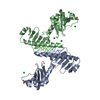
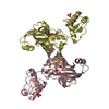
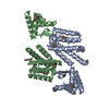
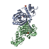
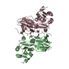
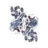
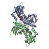
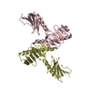
A: Chimera protein of Serine/threonine-protein kinase PLK4 and DDB1- and CUL4-associated factor 1 B: Chimera protein of Serine/threonine-protein kinase PLK4 and DDB1- and CUL4-associated factor 1
 Movie
Movie Controller

 Yorodumi
 Open data
 Basic information
 Components
 Keywords
 Function and homology information
 Homo sapiens (human)
 X-RAY DIFFRACTION /
  Authors
 United States, 1items
  Citation
 Structure visualization
 Downloads & links
 Other downloads
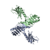
 PDBj
PDBj Assembly
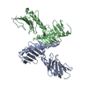

 Sample preparation
 Processing