Synthesis And Processing Of GAG, GAGPOL Polyproteins / host cellular component / host cell nuclear membrane / Integration of viral DNA into host genomic DNA / Autointegration results in viral DNA circles / Minus-strand DNA synthesis / Plus-strand DNA synthesis / Uncoating of the HIV Virion / 2-LTR circle formation / Vpr-mediated nuclear import of PICs ...Synthesis And Processing Of GAG, GAGPOL Polyproteins / host cellular component / host cell nuclear membrane / Integration of viral DNA into host genomic DNA / Autointegration results in viral DNA circles / Minus-strand DNA synthesis / Plus-strand DNA synthesis / Uncoating of the HIV Virion / 2-LTR circle formation / Vpr-mediated nuclear import of PICs / Early Phase of HIV Life Cycle / viral budding via host ESCRT complex / Integration of provirus / APOBEC3G mediated resistance to HIV-1 infection / Binding and entry of HIV virion / Membrane binding and targetting of GAG proteins / Assembly Of The HIV Virion / Budding and maturation of HIV virion / host multivesicular body / viral nucleocapsid / host cell cytoplasm / viral translational frameshifting / host cell nucleus / host cell plasma membrane / virion membrane / structural molecule activity / RNA binding / zinc ion binding Similarity search - Function
Retrovirus capsid C-terminal domain / Gag protein p6 / Gag protein p6 / Non-ribosomal Peptide Synthetase Peptidyl Carrier Protein; Chain A / : / gag protein p24 N-terminal domain / Immunodeficiency lentiviral matrix, N-terminal / gag gene protein p17 (matrix protein) / Matrix protein, lentiviral and alpha-retroviral, N-terminal / Retroviral nucleocapsid Gag protein p24, C-terminal domain ...Retrovirus capsid C-terminal domain / Gag protein p6 / Gag protein p6 / Non-ribosomal Peptide Synthetase Peptidyl Carrier Protein; Chain A / : / gag protein p24 N-terminal domain / Immunodeficiency lentiviral matrix, N-terminal / gag gene protein p17 (matrix protein) / Matrix protein, lentiviral and alpha-retroviral, N-terminal / Retroviral nucleocapsid Gag protein p24, C-terminal domain / Gag protein p24 C-terminal domain / Retrovirus capsid, C-terminal / Retroviral matrix protein / Retrovirus capsid, N-terminal / zinc finger / Zinc knuckle / Zinc finger, CCHC-type superfamily / Zinc finger, CCHC-type / Zinc finger CCHC-type profile. / Orthogonal Bundle / Mainly Alpha Similarity search - Domain/homology
National Institutes of Health/National Human Genome Research Institute (NIH/NHGRI)
5 P50 GM082545-10
United States
National Institutes of Health/National Institute of General Medical Sciences (NIH/NIGMS)
R01 GM066087
United States
National Institutes of Health/National Institute of General Medical Sciences (NIH/NIGMS)
P50 GM082545
United States
National Institutes of Health/National Institute of General Medical Sciences (NIH/NIGMS)
R01 GM12850
United States
Citation
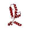
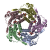
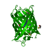
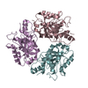
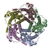
Journal: Proc Natl Acad Sci U S A / Year: 2018 Title: MicroED structures of HIV-1 Gag CTD-SP1 reveal binding interactions with the maturation inhibitor bevirimat. Authors: Michael D Purdy / Dan Shi / Jakub Chrustowicz / Johan Hattne / Tamir Gonen / Mark Yeager / Abstract: HIV-1 protease (PR) cleavage of the Gag polyprotein triggers the assembly of mature, infectious particles. Final cleavage of Gag occurs at the junction helix between the capsid protein CA and the SP1 ...HIV-1 protease (PR) cleavage of the Gag polyprotein triggers the assembly of mature, infectious particles. Final cleavage of Gag occurs at the junction helix between the capsid protein CA and the SP1 spacer peptide. Here we used MicroED to delineate the binding interactions of the maturation inhibitor bevirimat (BVM) using very thin frozen-hydrated, 3D microcrystals of a CTD-SP1 Gag construct with and without bound BVM. The 2.9-Å MicroED structure revealed that a single BVM molecule stabilizes the six-helix bundle via both electrostatic interactions with the dimethylsuccinyl moiety and hydrophobic interactions with the pentacyclic triterpenoid ring. These results provide insight into the mechanism of action of BVM and related maturation inhibitors that will inform further drug discovery efforts. This study also demonstrates the capabilities of MicroED for structure-based drug design.
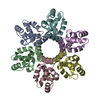
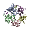
A: CTD-SP1 fragment of HIV-1 Gag B: CTD-SP1 fragment of HIV-1 Gag C: CTD-SP1 fragment of HIV-1 Gag D: CTD-SP1 fragment of HIV-1 Gag E: CTD-SP1 fragment of HIV-1 Gag F: CTD-SP1 fragment of HIV-1 Gag
Instrument: FEI VITROBOT MARK IV / Cryogen name: ETHANE
Crystal
Density Matthews: 2.31 Å3/Da / Density % sol: 46.81 %
Crystal grow
Temperature: 295 K / Method: vapor diffusion / pH: 7 / Details: 0.1 M Bis-Tris Propane, 1.1 M LiSO4
-
Data collection
Experimental equipment
Model: Tecnai F20 / Image courtesy: FEI Company
Microscopy
Model: FEI TECNAI F20
Electron gun
Electron source: FIELD EMISSION GUN / Accelerating voltage: 200 kV / Illumination mode: FLOOD BEAM
Electron lens
Mode: DIFFRACTION
Image recording
Average exposure time: 8 sec. / Electron dose: 0.05 e/Å2 / Film or detector model: TVIPS TEMCAM-F416 (4k x 4k) / Num. of grids imaged: 6 Details: Data from 6 crystals was merged for structure determination.
EM diffraction
Camera length: 2000 mm
EM diffraction shell
Resolution (Å)
ID
EM diffraction stats-ID
Fourier space coverage (%)
Multiplicity
Num. of structure factors
Phase residual (°)
3-19.9
1
1
79.8
5.7
10480
1
3-3.2
2
1
78.7
5.7
1006
1
EM diffraction stats
Fourier space coverage: 79.8 % / High resolution: 3 Å / Num. of intensities measured: 10480 / Num. of structure factors: 10480 / Phase error: 0 ° / Phase residual: 1 ° / Phase error rejection criteria: 0 / Rmerge: 0.564 / Rsym: 0.255
Diffraction source
Source: ELECTRON MICROSCOPE / Wavelength: 0.0251 Å
Radiation
Protocol: SINGLE WAVELENGTH / Monochromatic (M) / Laue (L): M / Scattering type: electron
Radiation wavelength
Wavelength: 0.0251 Å / Relative weight: 1
Reflection
Resolution: 3→27.5 Å / Num. obs: 10480 / % possible obs: 79.8 % / Redundancy: 5.7 % / Biso Wilson estimate: 55.82 Å2 / CC1/2: 0.9 / Rpim(I) all: 0.255 / Net I/σ(I): 3
Reflection shell
Resolution: 3→3.2 Å / Redundancy: 5.7 % / Mean I/σ(I) obs: 0.7 / Num. unique all: 1006 / CC1/2: 0.35 / Rpim(I) all: 1.324 / % possible all: 78.7
-
Phasing
Phasing
Method: molecular replacement
-
Processing
Software
Name
Version
Classification
NB
MOSFLM
datareduction
Aimless
datascaling
PHASER
phasing
PHENIX
dev_2747
refinement
PDB_EXTRACT
3.24
dataextraction
EM software
ID
Name
Version
Category
8
PHENIX
dev-2747
molecularreplacement
11
AIMLESS
crystallographymerging
13
PHENIX
dev-2747
modelrefinement
EM 3D crystal entity
∠α: 90 ° / ∠β: 90 ° / ∠γ: 96.55 ° / A: 70.61 Å / B: 122.55 Å / C: 78.7 Å / Space group name: C121 / Space group num: 5
CTF correction
Type: NONE
3D reconstruction
Resolution: 3 Å / Resolution method: DIFFRACTION PATTERN/LAYERLINES / Symmetry type: 3D CRYSTAL
Atomic model building
B value: 41.1 / Protocol: FLEXIBLE FIT / Space: RECIPROCAL
In the structure databanks used in Yorodumi, some data are registered as the other names, "COVID-19 virus" and "2019-nCoV". Here are the details of the virus and the list of structure data.
Jan 31, 2019. EMDB accession codes are about to change! (news from PDBe EMDB page)
EMDB accession codes are about to change! (news from PDBe EMDB page)
The allocation of 4 digits for EMDB accession codes will soon come to an end. Whilst these codes will remain in use, new EMDB accession codes will include an additional digit and will expand incrementally as the available range of codes is exhausted. The current 4-digit format prefixed with “EMD-” (i.e. EMD-XXXX) will advance to a 5-digit format (i.e. EMD-XXXXX), and so on. It is currently estimated that the 4-digit codes will be depleted around Spring 2019, at which point the 5-digit format will come into force.
The EM Navigator/Yorodumi systems omit the EMD- prefix.
Related info.:Q: What is EMD? / ID/Accession-code notation in Yorodumi/EM Navigator
Yorodumi is a browser for structure data from EMDB, PDB, SASBDB, etc.
This page is also the successor to EM Navigator detail page, and also detail information page/front-end page for Omokage search.
The word "yorodu" (or yorozu) is an old Japanese word meaning "ten thousand". "mi" (miru) is to see.
Related info.:EMDB / PDB / SASBDB / Comparison of 3 databanks / Yorodumi Search / Aug 31, 2016. New EM Navigator & Yorodumi / Yorodumi Papers / Jmol/JSmol / Function and homology information / Changes in new EM Navigator and Yorodumi
 Movie
Movie Controller


 Open data
 Basic information
 Components
 Keywords
 Function and homology information
 Human immunodeficiency virus 1
 MOLECULAR REPLACEMENT /
  Authors
 United States, 4items
  Citation
 Structure visualization
 Downloads & links
 Other downloads
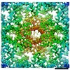


 PDBj
PDBj


 Assembly

 Sample preparation
 Processing