Resolution: 1.6→1.63 Å / Redundancy: 7.8 % / Rmerge(I) obs: 1.338 / Mean I/σ(I) obs: 1.51 / Num. unique obs: 1543 / CC1/2: 0.603 / % possible all: 93.6
-
Processing
Software
Name
Version
Classification
PHENIX
(1.14rc1_3161: ???)
refinement
HKL-3000
datareduction
HKL-3000
datascaling
HKL-3000
phasing
SBC-Collect
datacollection
Refinement
Method to determine structure: SAD / Resolution: 1.601→27.916 Å / SU ML: 0.17 / Cross valid method: THROUGHOUT / σ(F): 1.36 / Phase error: 19.76 / Stereochemistry target values: ML
Rfactor
Num. reflection
% reflection
Rfree
0.201
1645
5 %
Rwork
0.1598
-
-
obs
0.1619
32929
97.46 %
Solvent computation
Shrinkage radii: 0.9 Å / VDW probe radii: 1.11 Å / Solvent model: FLAT BULK SOLVENT MODEL
Refinement step
Cycle: LAST / Resolution: 1.601→27.916 Å
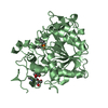
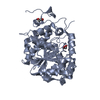
Protein
Nucleic acid
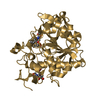
Ligand
Solvent
Total
Num. atoms
2096
0
16
274
2386
Refine LS restraints
Refine-ID
Type
Dev ideal
Number
X-RAY DIFFRACTION
f_bond_d
0.009
2208
X-RAY DIFFRACTION
f_angle_d
1.061
2975
X-RAY DIFFRACTION
f_dihedral_angle_d
13.969
1330
X-RAY DIFFRACTION
f_chiral_restr
0.061
304
X-RAY DIFFRACTION
f_plane_restr
0.006
377
LS refinement shell
Resolution (Å)
Rfactor Rfree
Num. reflection Rfree
Rfactor Rwork
Num. reflection Rwork
Refine-ID
% reflection obs (%)
1.6008-1.6479
0.2987
108
0.2586
2445
X-RAY DIFFRACTION
93
1.6479-1.7011
0.302
133
0.2328
2484
X-RAY DIFFRACTION
95
1.7011-1.7618
0.2317
143
0.2176
2522
X-RAY DIFFRACTION
95
1.7618-1.8324
0.2716
118
0.1998
2551
X-RAY DIFFRACTION
96
1.8324-1.9158
0.2156
141
0.1828
2527
X-RAY DIFFRACTION
96
1.9158-2.0167
0.2051
112
0.1629
2597
X-RAY DIFFRACTION
97
2.0167-2.143
0.1745
135
0.1446
2601
X-RAY DIFFRACTION
98
2.143-2.3084
0.1975
166
0.1461
2593
X-RAY DIFFRACTION
99
2.3084-2.5406
0.1797
147
0.1406
2688
X-RAY DIFFRACTION
100
2.5406-2.9079
0.1573
138
0.1539
2712
X-RAY DIFFRACTION
100
2.9079-3.6623
0.2117
147
0.1425
2710
X-RAY DIFFRACTION
100
3.6623-27.9205
0.1878
157
0.1471
2854
X-RAY DIFFRACTION
100
Refinement TLS params.
Method: refined / Refine-ID: X-RAY DIFFRACTION
ID
L11 (°2)
L12 (°2)
L13 (°2)
L22 (°2)
L23 (°2)
L33 (°2)
S11 (Å °)
S12 (Å °)
S13 (Å °)
S21 (Å °)
S22 (Å °)
S23 (Å °)
S31 (Å °)
S32 (Å °)
S33 (Å °)
T11 (Å2)
T12 (Å2)
T13 (Å2)
T22 (Å2)
T23 (Å2)
T33 (Å2)
Origin x (Å)
Origin y (Å)
Origin z (Å)
1
0.1197
-0.0479
0.012
0.108
0.1019
0.1215
0.0202
-0.0783
0.0913
-0.0231
-0.0203
0.0137
-0.1077
-0.0411
-0.0087
0.1311
0.0177
0.0104
0.0735
0.0047
0.1063
15.951
32.5349
34.7102
2
0.0177
-0.067
0.0468
0.3139
-0.112
0.5444
0.0338
0.0064
-0.0282
-0.0969
-0.001
0.0524
0.0628
0.0541
0.0473
0.0692
-0.0025
-0.0055
0.0584
-0.0016
0.0688
20.9696
8.4749
17.0219
3
0.1105
-0.0367
-0.0083
0.0587
0.0251
0.0056
0.0629
0.1201
-0.0093
-0.0369
-0.0744
0.0934
-0.0149
-0.0375
-0.0015
0.1069
-0.0369
-0.0074
0.1116
-0.0064
0.1325
9.097
5.8721
19.9824
4
0.105
-0.0191
-0.0228
0.0233
0.0344
0.0344
-0.0062
0.0018
-0.0057
-0.0084
0.0067
0.0375
0.0233
-0.0928
-0.006
0.0585
-0.0179
-0.0065
0.085
0.0035
0.075
8.5046
16.5152
23.842
5
0.0168
0.0104
0.0102
0.097
0.0649
0.0768
0.1034
-0.0924
-0.0304
0.0844
0.0012
-0.0065
0.1591
-0.029
0.0302
0.1343
-0.0289
-0.0052
0.0991
0.0228
0.0973
17.1202
5.322
34.8412
6
0.0662
0.0388
0.0412
0.0586
-0.0217
0.0281
0.0333
0.0216
-0.045
-0.0279
-0.0039
0.036
-0.0111
0.0651
0.0078
0.0683
-0.0041
-0.0041
0.111
0.0065
0.0799
22.8468
21.4576
25.7248
7
0.0513
-0.0106
-0.0299
0.0337
0.0285
0.0299
-0.0274
-0.0637
0.1015
0.0377
-0.0226
0.0221
-0.1129
-0.0635
0.0002
0.1028
0.0058
-0.0162
0.0978
-0.0076
0.0897
20.6166
26.5707
30.9952
8
0.0334
-0.0084
-0.0367
0.074
0.0042
0.0396
-0.0645
0.1025
-0.0213
0.0108
0.0033
-0.054
-0.133
0.1778
0
0.1267
-0.0273
-0.0104
0.1173
0.0081
0.106
26.5321
31.2062
24.6082
Refinement TLS group
ID
Refine-ID
Refine TLS-ID
Selection details
1
X-RAY DIFFRACTION
1
chain 'A' and (resid15through58 )
2
X-RAY DIFFRACTION
2
chain 'A' and (resid59through116 )
3
X-RAY DIFFRACTION
3
chain 'A' and (resid117through136 )
4
X-RAY DIFFRACTION
4
chain 'A' and (resid137through172 )
5
X-RAY DIFFRACTION
5
chain 'A' and (resid173through188 )
6
X-RAY DIFFRACTION
6
chain 'A' and (resid189through222 )
7
X-RAY DIFFRACTION
7
chain 'A' and (resid223through238 )
8
X-RAY DIFFRACTION
8
chain 'A' and (resid239through264 )
+
About Yorodumi
-
News
-
Feb 9, 2022. New format data for meta-information of EMDB entries
New format data for meta-information of EMDB entries
Version 3 of the EMDB header file is now the official format.
The previous official version 1.9 will be removed from the archive.
In the structure databanks used in Yorodumi, some data are registered as the other names, "COVID-19 virus" and "2019-nCoV". Here are the details of the virus and the list of structure data.
Jan 31, 2019. EMDB accession codes are about to change! (news from PDBe EMDB page)
EMDB accession codes are about to change! (news from PDBe EMDB page)
The allocation of 4 digits for EMDB accession codes will soon come to an end. Whilst these codes will remain in use, new EMDB accession codes will include an additional digit and will expand incrementally as the available range of codes is exhausted. The current 4-digit format prefixed with “EMD-” (i.e. EMD-XXXX) will advance to a 5-digit format (i.e. EMD-XXXXX), and so on. It is currently estimated that the 4-digit codes will be depleted around Spring 2019, at which point the 5-digit format will come into force.
The EM Navigator/Yorodumi systems omit the EMD- prefix.
Related info.:Q: What is EMD? / ID/Accession-code notation in Yorodumi/EM Navigator
Yorodumi is a browser for structure data from EMDB, PDB, SASBDB, etc.
This page is also the successor to EM Navigator detail page, and also detail information page/front-end page for Omokage search.
The word "yorodu" (or yorozu) is an old Japanese word meaning "ten thousand". "mi" (miru) is to see.
Related info.:EMDB / PDB / SASBDB / Comparison of 3 databanks / Yorodumi Search / Aug 31, 2016. New EM Navigator & Yorodumi / Yorodumi Papers / Jmol/JSmol / Function and homology information / Changes in new EM Navigator and Yorodumi
 Movie
Movie Controller

 Yorodumi
 Open data
 Basic information
 Components
 Keywords
 Function and homology information
 Sebaldella termitidis (bacteria)
 X-RAY DIFFRACTION /
  Authors
 United States, 1items
  Citation
 Structure visualization
 Downloads & links
 Other downloads
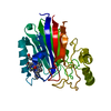

 PDBj
PDBj Assembly

 Mass: 96.063 Da / Num. of mol.: 2 / Source method: obtained synthetically / Formula: SO4
 Mass: 92.094 Da / Num. of mol.: 1 / Source method: obtained synthetically / Formula: C3H8O3
 Mass: 18.015 Da / Num. of mol.: 274 / Source method: isolated from a natural source / Formula: H2O
 Sample preparation
 Processing