 Movie
Movie Controller

[English] 日本語
 Yorodumi
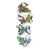
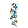
- PDB-6mxr: Crystal structure of the dimeric bH1-Fab variant [HC-Y33W,HC-D98M... -
+ Open data

- Basic information
| Entry | Database: PDB / ID: 6mxr | ||||||
|---|---|---|---|---|---|---|---|
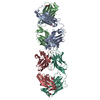
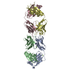
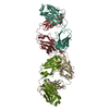
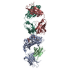
| Title | Crystal structure of the dimeric bH1-Fab variant [HC-Y33W,HC-D98M,HC-G99M] | ||||||
 Components Components | (anti-VEGF-A Fab fragment bH1 ...) x 2 | ||||||
 Keywords Keywords | IMMUNE SYSTEM / Fab fragment / antibody self-assembly / aggregation | ||||||
| Function / homology |  Function and homology information Function and homology informationimmunoglobulin complex / adaptive immune response / extracellular region / plasma membrane Similarity search - Function | ||||||
| Biological species |  Homo sapiens (human) Homo sapiens (human) | ||||||
| Method |  X-RAY DIFFRACTION / SYNCHROTRON / MOLECULAR REPLACEMENT / Resolution: 2.04 Å X-RAY DIFFRACTION / SYNCHROTRON / MOLECULAR REPLACEMENT / Resolution: 2.04 Å | ||||||
 Authors Authors | Shi, R. | ||||||
 Citation Citation | Journal: Mabs / Year: 2019 Title: Binding symmetry and surface flexibility mediate antibody self-association. Authors: Schrag, J.D. / Picard, M.E. / Gaudreault, F. / Gagnon, L.P. / Baardsnes, J. / Manenda, M.S. / Sheff, J. / Deprez, C. / Baptista, C. / Hogues, H. / Kelly, J.F. / Purisima, E.O. / Shi, R. / Sulea, T. | ||||||
| History |
|
- Structure visualization
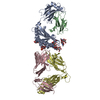
| Structure viewer | Molecule: MolmilJmol/JSmol |
|---|
- Downloads & links
-Download
| PDBx/mmCIF format | 6mxr.cif.gz | 199.7 KB | Display | PDBx/mmCIF format |
|---|---|---|---|---|
| PDB format | pdb6mxr.ent.gz | 154.8 KB | Display | PDB format |
| PDBx/mmJSON format | 6mxr.json.gz | Tree view | PDBx/mmJSON format | |
| Others |  Other downloads Other downloads |
-Validation report
| Arichive directory | https://data.pdbj.org/pub/pdb/validation_reports/mx/6mxrftp://data.pdbj.org/pub/pdb/validation_reports/mx/6mxr | HTTPS FTP |
|---|
-Related structure data
| Related structure data | 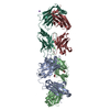 6mxsC  6my4C 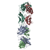 6my5C 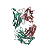 3bdyS C: citing same article ( S: Starting model for refinement |
|---|---|
| Similar structure data |


-Links
 PDBj
PDBj

- Assembly
| Deposited unit | 
| ||||||||
|---|---|---|---|---|---|---|---|---|---|
| 1 |
| ||||||||
| Unit cell |
|
-Components
-Antibody , 2 types, 4 molecules HALB
| #1: Antibody | Mass: 25422.518 Da / Num. of mol.: 2 / Mutation: Y33W, D98M, G99M Source method: isolated from a genetically manipulated source Source: (gene. exp.) Homo sapiens (human) / Cell line (production host): CHO-3E7 / Production host:   Cricetulus griseus (Chinese hamster) / References: UniProt: V9HW68 Cricetulus griseus (Chinese hamster) / References: UniProt: V9HW68#2: Antibody | Mass: 23956.588 Da / Num. of mol.: 2 Source method: isolated from a genetically manipulated source Source: (gene. exp.) Homo sapiens (human) / Cell line (production host): CHO-3E7 / Production host: Cricetulus griseus (Chinese hamster) / References: UniProt: Q7Z3Y4 |
|---|
-Non-polymers , 4 types, 726 molecules 


| #3: Chemical |  Mass: 35.453 Da / Num. of mol.: 2 / Source method: obtained synthetically / Formula: Cl Mass: 35.453 Da / Num. of mol.: 2 / Source method: obtained synthetically / Formula: Cl#4: Chemical | ChemComp-NA / |  Mass: 22.990 Da / Num. of mol.: 1 / Source method: obtained synthetically / Formula: Na Mass: 22.990 Da / Num. of mol.: 1 / Source method: obtained synthetically / Formula: Na#5: Chemical | ChemComp-EDO / |  Mass: 62.068 Da / Num. of mol.: 1 / Source method: obtained synthetically / Formula: C2H6O2 Mass: 62.068 Da / Num. of mol.: 1 / Source method: obtained synthetically / Formula: C2H6O2#6: Water | ChemComp-HOH / | Mass: 18.015 Da / Num. of mol.: 722 / Source method: isolated from a natural source / Formula: H2O |
|---|
-Details
| Has protein modification | Y |
|---|
-Experimental details
-Experiment
| Experiment | Method: X-RAY DIFFRACTION / Number of used crystals: 1 |
|---|
- Sample preparation
| Crystal | Density Matthews: 3.38 Å3/Da / Density % sol: 63.59 % |
|---|---|
| Crystal grow | Temperature: 295 K / Method: microbatch / pH: 5.6 Details: 0.1 M tris-sodium citrate, pH 5.6, 11% PEG4000, 8% isopropanol |
-Data collection
| Diffraction | Mean temperature: 100 K / Serial crystal experiment: N | ||||||||||||||||||||||||
|---|---|---|---|---|---|---|---|---|---|---|---|---|---|---|---|---|---|---|---|---|---|---|---|---|---|
| Diffraction source | Source: SYNCHROTRON / Site: CLSI  / Beamline: 08ID-1 / Wavelength: 0.9795 Å / Beamline: 08ID-1 / Wavelength: 0.9795 Å | ||||||||||||||||||||||||
| Detector | Type: RAYONIX MX-300 / Detector: CCD / Date: Jan 7, 2017 | ||||||||||||||||||||||||
| Radiation | Monochromator: double crystal Si(111) / Protocol: SINGLE WAVELENGTH / Monochromatic (M) / Laue (L): M / Scattering type: x-ray | ||||||||||||||||||||||||
| Radiation wavelength | Wavelength: 0.9795 Å / Relative weight: 1 | ||||||||||||||||||||||||
| Reflection | Resolution: 2.04→50 Å / Num. obs: 83010 / % possible obs: 99.8 % / Redundancy: 3.4 % / CC1/2: 0.995 / Rmerge(I) obs: 0.087 / Rpim(I) all: 0.055 / Rrim(I) all: 0.104 / Net I/σ(I): 8.3 | ||||||||||||||||||||||||
| Reflection shell | Diffraction-ID: 1
|
- Processing
| Software |
| ||||||||||||||||||||||||||||||||||||||||||||||||||||||||||||
|---|---|---|---|---|---|---|---|---|---|---|---|---|---|---|---|---|---|---|---|---|---|---|---|---|---|---|---|---|---|---|---|---|---|---|---|---|---|---|---|---|---|---|---|---|---|---|---|---|---|---|---|---|---|---|---|---|---|---|---|---|---|
| Refinement | Method to determine structure: MOLECULAR REPLACEMENT Starting model: PDB entry 3BDY Resolution: 2.04→50 Å / Cor.coef. Fo:Fc: 0.965 / Cor.coef. Fo:Fc free: 0.952 / SU B: 4.045 / SU ML: 0.104 / Cross valid method: THROUGHOUT / σ(F): 0 / ESU R: 0.135 / ESU R Free: 0.128 Details: HYDROGENS HAVE BEEN ADDED IN THE RIDING POSITIONS U VALUES : REFINED INDIVIDUALLY
| ||||||||||||||||||||||||||||||||||||||||||||||||||||||||||||
| Solvent computation | Ion probe radii: 0.8 Å / Shrinkage radii: 0.8 Å / VDW probe radii: 1.2 Å | ||||||||||||||||||||||||||||||||||||||||||||||||||||||||||||
| Displacement parameters | Biso max: 107.54 Å2 / Biso mean: 32.147 Å2 / Biso min: 13.52 Å2
| ||||||||||||||||||||||||||||||||||||||||||||||||||||||||||||
| Refinement step | Cycle: final / Resolution: 2.04→50 Å
| ||||||||||||||||||||||||||||||||||||||||||||||||||||||||||||
| Refine LS restraints |
| ||||||||||||||||||||||||||||||||||||||||||||||||||||||||||||
| LS refinement shell | Resolution: 2.04→2.093 Å / Rfactor Rfree error: 0 / Total num. of bins used: 20
|
