 Movie
Movie Controller

[English] 日本語
 Yorodumi
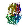
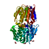
- PDB-6mm7: Catalytic subunit of cAMP-dependent protein kinase A in complex w... -
+ Open data

- Basic information
| Entry | Database: PDB / ID: 6mm7 | ||||||
|---|---|---|---|---|---|---|---|
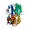
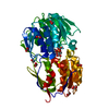
| Title | Catalytic subunit of cAMP-dependent protein kinase A in complex with RyR2 K2879A, S2813D phosphomimetic (2699-2904) crystal form 1 | ||||||
 Components Components |
| ||||||
 Keywords Keywords | TRANSFERASE/PROTEIN BINDING / Kinase / complex / ion channel / enzyme / PROTEIN BINDING / TRANSFERASE-PROTEIN BINDING complex | ||||||
| Function / homology |  Function and homology information Function and homology informationmanganese ion transmembrane transport / spontaneous exocytosis of neurotransmitter / PKA activation in glucagon signalling / CREB1 phosphorylation through the activation of Adenylate Cyclase / negative regulation of meiotic cell cycle / HDL assembly / positive regulation of triglyceride catabolic process / establishment of protein localization to endoplasmic reticulum / : / DARPP-32 events ...manganese ion transmembrane transport / spontaneous exocytosis of neurotransmitter / PKA activation in glucagon signalling / CREB1 phosphorylation through the activation of Adenylate Cyclase / negative regulation of meiotic cell cycle / HDL assembly / positive regulation of triglyceride catabolic process / establishment of protein localization to endoplasmic reticulum / : / DARPP-32 events / type B pancreatic cell apoptotic process / Rap1 signalling / Purkinje myocyte to ventricular cardiac muscle cell signaling / regulation of atrial cardiac muscle cell action potential / PKA activation / Regulation of insulin secretion / left ventricular cardiac muscle tissue morphogenesis / suramin binding / Vasopressin regulates renal water homeostasis via Aquaporins / regulation of AV node cell action potential / GPER1 signaling / Glucagon-like Peptide-1 (GLP1) regulates insulin secretion / Hedgehog 'off' state / Loss of Nlp from mitotic centrosomes / Recruitment of mitotic centrosome proteins and complexes / Loss of proteins required for interphase microtubule organization from the centrosome / sarcoplasmic reticulum calcium ion transport / Anchoring of the basal body to the plasma membrane / Recruitment of NuMA to mitotic centrosomes / regulation of SA node cell action potential / AURKA Activation by TPX2 / Factors involved in megakaryocyte development and platelet production / MAPK6/MAPK4 signaling / Interleukin-3, Interleukin-5 and GM-CSF signaling / CD209 (DC-SIGN) signaling / RET signaling / GLI3 is processed to GLI3R by the proteasome / High laminar flow shear stress activates signaling by PIEZO1 and PECAM1:CDH5:KDR in endothelial cells / calcium-induced calcium release activity / A band / regulation of cellular respiration / Regulation of PLK1 Activity at G2/M Transition / Stimuli-sensing channels / Mitochondrial protein degradation / VEGFA-VEGFR2 Pathway / ventricular cardiac muscle cell action potential / nucleotide-activated protein kinase complex / Ion homeostasis / cardiac muscle hypertrophy / embryonic heart tube morphogenesis / regulation of ventricular cardiac muscle cell action potential / calcium ion transport into cytosol / ryanodine-sensitive calcium-release channel activity / potassium channel inhibitor activity / histone H1-4S35 kinase activity / cAMP-dependent protein kinase / release of sequestered calcium ion into cytosol by sarcoplasmic reticulum / negative regulation of glycolytic process / regulation of protein processing / response to redox state / cAMP-dependent protein kinase activity / regulation of cardiac muscle contraction by calcium ion signaling / cellular response to parathyroid hormone stimulus / protein localization to lipid droplet / cellular response to caffeine / regulation of bicellular tight junction assembly / cAMP-dependent protein kinase complex / extrinsic component of cytoplasmic side of plasma membrane / response to caffeine / response to muscle activity / interleukin-2-mediated signaling pathway / sperm capacitation / regulation of osteoblast differentiation / peptidyl-threonine phosphorylation / calcium ion transmembrane import into cytosol / cellular response to cold / protein kinase A regulatory subunit binding / negative regulation of glycolytic process through fructose-6-phosphate / ciliary base / protein kinase A catalytic subunit binding / negative regulation of cytosolic calcium ion concentration / positive regulation of the force of heart contraction / smooth endoplasmic reticulum / intracellularly gated calcium channel activity / intracellular potassium ion homeostasis / TORC1 signaling / mesoderm formation / response to magnesium ion / plasma membrane raft / cAMP/PKA signal transduction / axoneme / detection of calcium ion / regulation of cardiac muscle contraction / sperm flagellum / postsynaptic modulation of chemical synaptic transmission / regulation of cytosolic calcium ion concentration / positive regulation of heart rate / regulation of synaptic transmission, glutamatergic / release of sequestered calcium ion into cytosol / response to muscle stretch Similarity search - Function | ||||||
| Biological species |  | ||||||
| Method |  X-RAY DIFFRACTION / SYNCHROTRON / MOLECULAR REPLACEMENT / Resolution: 1.85 Å X-RAY DIFFRACTION / SYNCHROTRON / MOLECULAR REPLACEMENT / Resolution: 1.85 Å | ||||||
 Authors Authors | van Petegem, F. / Haji-Ghassemi, O. | ||||||
| Funding support |  Canada, 1items Canada, 1items
| ||||||
 Citation Citation | Journal: Mol.Cell / Year: 2019 Title: cAMP-dependent protein kinase A in complex with RyR2 peptide (2799-2810) Authors: Haji-Ghassemi, O. / Yuchi, Z. / van Petegem, F. | ||||||
| History |
|
- Structure visualization
| Structure viewer | Molecule: MolmilJmol/JSmol |
|---|
- Downloads & links
-Download
| PDBx/mmCIF format | 6mm7.cif.gz | 466.8 KB | Display | PDBx/mmCIF format |
|---|---|---|---|---|
| PDB format | pdb6mm7.ent.gz | 377.9 KB | Display | PDB format |
| PDBx/mmJSON format | 6mm7.json.gz | Tree view | PDBx/mmJSON format | |
| Others |  Other downloads Other downloads |
-Validation report
| Arichive directory | https://data.pdbj.org/pub/pdb/validation_reports/mm/6mm7ftp://data.pdbj.org/pub/pdb/validation_reports/mm/6mm7 | HTTPS FTP |
|---|
-Related structure data
| Related structure data | 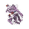 6mm5C  6mm6SC 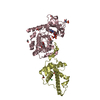 6mm8C S: Starting model for refinement C: citing same article ( |
|---|---|
| Similar structure data |


-Links
 PDBj
PDBj


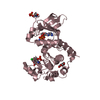
- Assembly
| Deposited unit | 
| ||||||||
|---|---|---|---|---|---|---|---|---|---|
| 1 | 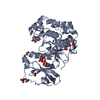
| ||||||||
| 2 | 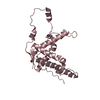
| ||||||||
| 3 | 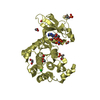
| ||||||||
| 4 | 
| ||||||||
| Unit cell |
|
-Components
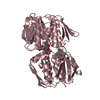
-Protein , 2 types, 4 molecules ADCE
| #1: Protein | Mass: 39598.191 Da / Num. of mol.: 2 / Fragment: residues 16-351 Source method: isolated from a genetically manipulated source Source: (gene. exp.)  #2: Protein | Mass: 24285.613 Da / Num. of mol.: 2 / Fragment: residues 2699-2904 / Mutation: K2879A, S2813D Source method: isolated from a genetically manipulated source Source: (gene. exp.) |
|---|
-Non-polymers , 6 types, 850 molecules 


| #3: Chemical | ChemComp-FMT /  Mass: 46.025 Da / Num. of mol.: 8 / Source method: obtained synthetically / Formula: CH2O2 Mass: 46.025 Da / Num. of mol.: 8 / Source method: obtained synthetically / Formula: CH2O2#4: Chemical | ChemComp-ACY / |  Mass: 60.052 Da / Num. of mol.: 1 / Source method: obtained synthetically / Formula: C2H4O2 Mass: 60.052 Da / Num. of mol.: 1 / Source method: obtained synthetically / Formula: C2H4O2#5: Chemical |  Mass: 506.196 Da / Num. of mol.: 2 / Source method: obtained synthetically / Formula: C10H17N6O12P3 / Feature type: SUBJECT OF INVESTIGATION / Comment: AMP-PNP, energy-carrying molecule analogue*YM Mass: 506.196 Da / Num. of mol.: 2 / Source method: obtained synthetically / Formula: C10H17N6O12P3 / Feature type: SUBJECT OF INVESTIGATION / Comment: AMP-PNP, energy-carrying molecule analogue*YM#6: Chemical | ChemComp-EDO /  Mass: 62.068 Da / Num. of mol.: 7 / Source method: obtained synthetically / Formula: C2H6O2 Mass: 62.068 Da / Num. of mol.: 7 / Source method: obtained synthetically / Formula: C2H6O2#7: Chemical |  Mass: 35.453 Da / Num. of mol.: 2 / Source method: obtained synthetically / Formula: Cl Mass: 35.453 Da / Num. of mol.: 2 / Source method: obtained synthetically / Formula: Cl#8: Water | ChemComp-HOH / | Mass: 18.015 Da / Num. of mol.: 830 / Source method: isolated from a natural source / Formula: H2O |
|---|
-Details
| Has protein modification | Y |
|---|
-Experimental details
-Experiment
| Experiment | Method: X-RAY DIFFRACTION / Number of used crystals: 1 |
|---|
- Sample preparation
| Crystal | Density Matthews: 2.73 Å3/Da / Density % sol: 54.92 % |
|---|---|
| Crystal grow | Temperature: 277 K / Method: vapor diffusion, sitting drop / pH: 7.5 Details: 0.1 M magnesium formate and 25% (w/v) PEG 3350; 0.05 M HEPES, and 25% ethylene glycol |
-Data collection
| Diffraction | Mean temperature: 100 K / Serial crystal experiment: N | ||||||||||||||||||||||||||||||||||||||||||||||||||||||||||||||||||||||||||||||||||||||||||||||||||||||||||||||||||||||||||||||||||||||||||||||||
|---|---|---|---|---|---|---|---|---|---|---|---|---|---|---|---|---|---|---|---|---|---|---|---|---|---|---|---|---|---|---|---|---|---|---|---|---|---|---|---|---|---|---|---|---|---|---|---|---|---|---|---|---|---|---|---|---|---|---|---|---|---|---|---|---|---|---|---|---|---|---|---|---|---|---|---|---|---|---|---|---|---|---|---|---|---|---|---|---|---|---|---|---|---|---|---|---|---|---|---|---|---|---|---|---|---|---|---|---|---|---|---|---|---|---|---|---|---|---|---|---|---|---|---|---|---|---|---|---|---|---|---|---|---|---|---|---|---|---|---|---|---|---|---|---|---|
| Diffraction source | Source: SYNCHROTRON / Site: CLSI / Beamline: 08B1-1 / Wavelength: 0.979 Å | ||||||||||||||||||||||||||||||||||||||||||||||||||||||||||||||||||||||||||||||||||||||||||||||||||||||||||||||||||||||||||||||||||||||||||||||||
| Detector | Type: MARMOSAIC 300 mm CCD / Detector: CCD / Date: Feb 8, 2018 | ||||||||||||||||||||||||||||||||||||||||||||||||||||||||||||||||||||||||||||||||||||||||||||||||||||||||||||||||||||||||||||||||||||||||||||||||
| Radiation | Protocol: SINGLE WAVELENGTH / Monochromatic (M) / Laue (L): M / Scattering type: x-ray | ||||||||||||||||||||||||||||||||||||||||||||||||||||||||||||||||||||||||||||||||||||||||||||||||||||||||||||||||||||||||||||||||||||||||||||||||
| Radiation wavelength | Wavelength: 0.979 Å / Relative weight: 1 | ||||||||||||||||||||||||||||||||||||||||||||||||||||||||||||||||||||||||||||||||||||||||||||||||||||||||||||||||||||||||||||||||||||||||||||||||
| Reflection | Resolution: 1.85→35 Å / Num. obs: 111231 / % possible obs: 94.4 % / Redundancy: 6.3 % / Rmerge(I) obs: 0.066 / Rpim(I) all: 0.027 / Rrim(I) all: 0.071 / Χ2: 1.059 / Net I/σ(I): 11.3 / Num. measured all: 698657 | ||||||||||||||||||||||||||||||||||||||||||||||||||||||||||||||||||||||||||||||||||||||||||||||||||||||||||||||||||||||||||||||||||||||||||||||||
| Reflection shell | Diffraction-ID: 1
|
- Processing
| Software |
| |||||||||||||||||||||||||||||||||||||||||||||||||||||||||||||||||||||||||||||||||||||||||||||||||||||||||||||||||||||||||||||
|---|---|---|---|---|---|---|---|---|---|---|---|---|---|---|---|---|---|---|---|---|---|---|---|---|---|---|---|---|---|---|---|---|---|---|---|---|---|---|---|---|---|---|---|---|---|---|---|---|---|---|---|---|---|---|---|---|---|---|---|---|---|---|---|---|---|---|---|---|---|---|---|---|---|---|---|---|---|---|---|---|---|---|---|---|---|---|---|---|---|---|---|---|---|---|---|---|---|---|---|---|---|---|---|---|---|---|---|---|---|---|---|---|---|---|---|---|---|---|---|---|---|---|---|---|---|---|
| Refinement | Method to determine structure: MOLECULAR REPLACEMENT Starting model: 6MM6 Resolution: 1.85→33.8 Å / Cor.coef. Fo:Fc: 0.951 / Cor.coef. Fo:Fc free: 0.944 / SU B: 5.157 / SU ML: 0.088 / Cross valid method: THROUGHOUT / σ(F): 0 / ESU R: 0.155 / ESU R Free: 0.139 Details: HYDROGENS HAVE BEEN ADDED IN THE RIDING POSITIONS U VALUES : WITH TLS ADDED
| |||||||||||||||||||||||||||||||||||||||||||||||||||||||||||||||||||||||||||||||||||||||||||||||||||||||||||||||||||||||||||||
| Solvent computation | Ion probe radii: 0.8 Å / Shrinkage radii: 0.8 Å / VDW probe radii: 1.2 Å | |||||||||||||||||||||||||||||||||||||||||||||||||||||||||||||||||||||||||||||||||||||||||||||||||||||||||||||||||||||||||||||
| Displacement parameters | Biso max: 128.62 Å2 / Biso mean: 37.297 Å2 / Biso min: 19.16 Å2
| |||||||||||||||||||||||||||||||||||||||||||||||||||||||||||||||||||||||||||||||||||||||||||||||||||||||||||||||||||||||||||||
| Refinement step | Cycle: final / Resolution: 1.85→33.8 Å
| |||||||||||||||||||||||||||||||||||||||||||||||||||||||||||||||||||||||||||||||||||||||||||||||||||||||||||||||||||||||||||||
| Refine LS restraints |
| |||||||||||||||||||||||||||||||||||||||||||||||||||||||||||||||||||||||||||||||||||||||||||||||||||||||||||||||||||||||||||||
| LS refinement shell | Resolution: 1.849→1.897 Å / Rfactor Rfree error: 0 / Total num. of bins used: 20
| |||||||||||||||||||||||||||||||||||||||||||||||||||||||||||||||||||||||||||||||||||||||||||||||||||||||||||||||||||||||||||||
| Refinement TLS params. | Method: refined / Refine-ID: X-RAY DIFFRACTION
| |||||||||||||||||||||||||||||||||||||||||||||||||||||||||||||||||||||||||||||||||||||||||||||||||||||||||||||||||||||||||||||
| Refinement TLS group |
|
