 Movie
Movie Controller

[English] 日本語
 Yorodumi
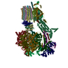
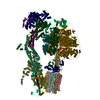
- PDB-6mem: A unique supramolecular organization of photosystem I in the moss... -
+ Open data

- Basic information
| Entry | Database: PDB / ID: 6mem | |||||||||
|---|---|---|---|---|---|---|---|---|---|---|
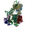
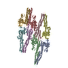
| Title | A unique supramolecular organization of photosystem I in the moss Physcomitrella patens | |||||||||
 Components Components |
| |||||||||
 Keywords Keywords | PHOTOSYNTHESIS / complex / photosystem I / light harvesting complex / Physcomitrella | |||||||||
| Biological species |  Physcomitrella patens (plant) Physcomitrella patens (plant) | |||||||||
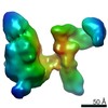
| Method | ELECTRON MICROSCOPY / single particle reconstruction / cryo EM / Resolution: 11.6 Å | |||||||||
 Authors Authors | Iwai, M. / Grob, P. | |||||||||
| Funding support |  United States, 2items United States, 2items
| |||||||||
 Citation Citation | Journal: Nat Plants / Year: 2018 Title: A unique supramolecular organization of photosystem I in the moss Physcomitrella patens. Authors: Masakazu Iwai / Patricia Grob / Anthony T Iavarone / Eva Nogales / Krishna K Niyogi / Abstract: The photosynthesis machinery in chloroplast thylakoid membranes is comprised of multiple protein complexes and supercomplexes. Here, we show a novel supramolecular organization of photosystem I (PSI) ...The photosynthesis machinery in chloroplast thylakoid membranes is comprised of multiple protein complexes and supercomplexes. Here, we show a novel supramolecular organization of photosystem I (PSI) in the moss Physcomitrella patens by single-particle cryo-electron microscopy. The moss-specific light-harvesting complex (LHC) protein Lhcb9 is involved in this PSI supercomplex, which has been shown to have a molecular density similar to that of the green alga Chlamydomonas reinhardtii. Our results show that the structural organization is unexpectedly different-two rows of the LHCI belt exist as in C. reinhardtii, but the outer one is shifted toward the PsaK side. Furthermore, one trimeric LHC protein and one monomeric LHC protein position alongside PsaL/K, filling the gap between these subunits and the outer LHCI belt. We provide evidence showing that Lhcb9 is a key factor, acting as a linkage between the PSI core and the outer LHCI belt to form the unique supramolecular organization of the PSI supercomplex in P. patens. | |||||||||
| History |
|
- Structure visualization
| Movie |
 Movie viewer Movie viewer |
|---|---|
| Structure viewer | Molecule: MolmilJmol/JSmol |
- Downloads & links
-Download
| PDBx/mmCIF format | 6mem.cif.gz | 666.8 KB | Display | PDBx/mmCIF format |
|---|---|---|---|---|
| PDB format | pdb6mem.ent.gz | 555.1 KB | Display | PDB format |
| PDBx/mmJSON format | 6mem.json.gz | Tree view | PDBx/mmJSON format | |
| Others |  Other downloads Other downloads |
-Validation report
| Arichive directory | https://data.pdbj.org/pub/pdb/validation_reports/me/6memftp://data.pdbj.org/pub/pdb/validation_reports/me/6mem | HTTPS FTP |
|---|
-Related structure data
| Related structure data | 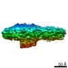 9107MC M: map data used to model this data C: citing same article ( |
|---|---|
| Similar structure data |


-Links
 PDBj
PDBj
- Assembly
| Deposited unit | 
|
|---|---|
| 1 |
|
-Components
-Chlorophyll A/B binding protein ... , 12 types, 12 molecules ABCDEFGHIKMO
| #1: Protein | Mass: 16443.229 Da / Num. of mol.: 1 / Source method: isolated from a natural source / Source: (natural) Physcomitrella patens (plant) |
|---|---|
| #2: Protein | Mass: 16613.436 Da / Num. of mol.: 1 / Source method: isolated from a natural source / Source: (natural) Physcomitrella patens (plant) |
| #3: Protein | Mass: 17719.787 Da / Num. of mol.: 1 / Source method: isolated from a natural source / Source: (natural) Physcomitrella patens (plant) |
| #4: Protein | Mass: 17549.580 Da / Num. of mol.: 1 / Source method: isolated from a natural source / Source: (natural) Physcomitrella patens (plant) |
| #5: Protein | Mass: 18826.139 Da / Num. of mol.: 1 / Source method: isolated from a natural source / Source: (natural) Physcomitrella patens (plant) |
| #6: Protein | Mass: 18570.826 Da / Num. of mol.: 1 / Source method: isolated from a natural source / Source: (natural) Physcomitrella patens (plant) |
| #7: Protein | Mass: 18655.930 Da / Num. of mol.: 1 / Source method: isolated from a natural source / Source: (natural) Physcomitrella patens (plant) |
| #8: Protein | Mass: 16868.748 Da / Num. of mol.: 1 / Source method: isolated from a natural source / Source: (natural) Physcomitrella patens (plant) |
| #9: Protein | Mass: 16698.539 Da / Num. of mol.: 1 / Source method: isolated from a natural source / Source: (natural) Physcomitrella patens (plant) |
| #11: Protein | Mass: 18996.346 Da / Num. of mol.: 1 / Source method: isolated from a natural source / Source: (natural) Physcomitrella patens (plant) |
| #13: Protein | Mass: 19081.451 Da / Num. of mol.: 1 / Source method: isolated from a natural source / Source: (natural) Physcomitrella patens (plant) |
| #15: Protein | Mass: 19166.555 Da / Num. of mol.: 1 / Source method: isolated from a natural source / Source: (natural) Physcomitrella patens (plant) |
-Protein , 10 types, 10 molecules JLNPQRSTWX
| #10: Protein | Mass: 63250.809 Da / Num. of mol.: 1 / Source method: isolated from a natural source / Source: (natural) Physcomitrella patens (plant) |
|---|---|
| #12: Protein | Mass: 62399.789 Da / Num. of mol.: 1 / Source method: isolated from a natural source / Source: (natural) Physcomitrella patens (plant) |
| #14: Protein | Mass: 6826.406 Da / Num. of mol.: 1 / Source method: isolated from a natural source / Source: (natural) Physcomitrella patens (plant) |
| #16: Protein | Mass: 12188.016 Da / Num. of mol.: 1 / Source method: isolated from a natural source / Source: (natural) Physcomitrella patens (plant) |
| #17: Protein | Mass: 5634.938 Da / Num. of mol.: 1 / Source method: isolated from a natural source / Source: (natural) Physcomitrella patens (plant) |
| #18: Protein | Mass: 13124.170 Da / Num. of mol.: 1 / Source method: isolated from a natural source / Source: (natural) Physcomitrella patens (plant) |
| #19: Protein | Mass: 8273.189 Da / Num. of mol.: 1 / Source method: isolated from a natural source / Source: (natural) Physcomitrella patens (plant) |
| #20: Protein | Mass: 7507.245 Da / Num. of mol.: 1 / Source method: isolated from a natural source / Source: (natural) Physcomitrella patens (plant) |
| #23: Protein | Mass: 6571.091 Da / Num. of mol.: 1 / Source method: isolated from a natural source / Source: (natural) Physcomitrella patens (plant) |
| #24: Protein | Mass: 13379.484 Da / Num. of mol.: 1 / Source method: isolated from a natural source / Source: (natural) Physcomitrella patens (plant) |
-Protein/peptide , 2 types, 2 molecules UV
| #21: Protein/peptide | Mass: 2571.161 Da / Num. of mol.: 1 / Source method: isolated from a natural source / Source: (natural) Physcomitrella patens (plant) |
|---|---|
| #22: Protein/peptide | Mass: 3592.419 Da / Num. of mol.: 1 / Source method: isolated from a natural source / Source: (natural) Physcomitrella patens (plant) |
-Experimental details
-Experiment
| Experiment | Method: ELECTRON MICROSCOPY |
|---|---|
| EM experiment | Aggregation state: PARTICLE / 3D reconstruction method: single particle reconstruction |
- Sample preparation
| Component | Name: Large PSI-LHCI supercomplex / Type: COMPLEX Details: A protein supercomplex isolated by gentle detergent solubilization of thylakoid membranes and maltose density gradient centrifugation Entity ID: #1-#6, #8-#12, #14, #16-#24, #7, #13, #15 / Source: NATURAL | ||||||||||||||||
|---|---|---|---|---|---|---|---|---|---|---|---|---|---|---|---|---|---|
| Molecular weight | Value: 0.94 MDa / Experimental value: NO | ||||||||||||||||
| Source (natural) | Organism: Physcomitrella patens (plant) / Strain: Gransden 2004 / Cellular location: chloroplast / Organelle: chloroplast / Tissue: protonema | ||||||||||||||||
| Buffer solution | pH: 6.5 | ||||||||||||||||
| Buffer component |
| ||||||||||||||||
| Specimen | Conc.: 0.2 mg/ml / Embedding applied: NO / Shadowing applied: NO / Staining applied: NO / Vitrification applied: YES | ||||||||||||||||
| Vitrification | Instrument: FEI VITROBOT MARK IV / Cryogen name: ETHANE / Humidity: 100 % / Chamber temperature: 283 K / Details: Lights off, blot 4.5 seconds before plunging. |
- Electron microscopy imaging
| Experimental equipment |  Model: Tecnai F20 / Image courtesy: FEI Company |
|---|---|
| Microscopy | Model: FEI TECNAI F20 |
| Electron gun | Electron source:  FIELD EMISSION GUN / Accelerating voltage: 120 kV / Illumination mode: FLOOD BEAM FIELD EMISSION GUN / Accelerating voltage: 120 kV / Illumination mode: FLOOD BEAM |
| Electron lens | Mode: BRIGHT FIELD / Nominal magnification: 80000 X / Calibrated magnification: 107140 X / Nominal defocus max: -1800 nm / Nominal defocus min: -2500 nm / Calibrated defocus min: -1900 nm / Calibrated defocus max: -4200 nm / Cs: 2.2 mm / C2 aperture diameter: 70 µm / Alignment procedure: COMA FREE |
| Specimen holder | Cryogen: NITROGEN Specimen holder model: GATAN 626 SINGLE TILT LIQUID NITROGEN CRYO TRANSFER HOLDER Temperature (max): 93 K / Temperature (min): 93 K |
| Image recording | Average exposure time: 0.5 sec. / Electron dose: 25 e/Å2 / Film or detector model: GATAN ULTRASCAN 4000 (4k x 4k) / Num. of grids imaged: 3 / Num. of real images: 361 |
| Image scans | Sampling size: 15 µm / Width: 4096 / Height: 4096 |
- Processing
| EM software |
| ||||||||||||||||||||||||||||||||||||||||
|---|---|---|---|---|---|---|---|---|---|---|---|---|---|---|---|---|---|---|---|---|---|---|---|---|---|---|---|---|---|---|---|---|---|---|---|---|---|---|---|---|---|
| CTF correction | Type: PHASE FLIPPING AND AMPLITUDE CORRECTION | ||||||||||||||||||||||||||||||||||||||||
| Particle selection | Num. of particles selected: 55780 | ||||||||||||||||||||||||||||||||||||||||
| Symmetry | Point symmetry: C1 (asymmetric) | ||||||||||||||||||||||||||||||||||||||||
| 3D reconstruction | Resolution: 11.6 Å / Resolution method: FSC 0.143 CUT-OFF / Num. of particles: 14412 / Algorithm: FOURIER SPACE / Num. of class averages: 5 / Symmetry type: POINT | ||||||||||||||||||||||||||||||||||||||||
| Atomic model building | Protocol: RIGID BODY FIT | ||||||||||||||||||||||||||||||||||||||||
| Refinement | Highest resolution: 11.6 Å |
