 Movie
Movie Controller

[English] 日本語
 Yorodumi
- PDB-6m2i: Structure of the 2-Aminoisobutyric acid Monooxygenase Hydroxylase -
+ Open data

- Basic information
| Entry | Database: PDB / ID: 6m2i | ||||||
|---|---|---|---|---|---|---|---|
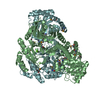
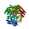
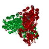
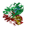
| Title | Structure of the 2-Aminoisobutyric acid Monooxygenase Hydroxylase | ||||||
 Components Components | (Amidohydrolase) x 2 | ||||||
 Keywords Keywords | OXIDOREDUCTASE / Monooxygenase / Hydroxylase | ||||||
| Function / homology |  Function and homology information Function and homology informationsecondary metabolic process / carboxy-lyase activity / hydrolase activity / metal ion binding / cytoplasm Similarity search - Function | ||||||
| Biological species |  Rhodococcus wratislaviensis (bacteria) Rhodococcus wratislaviensis (bacteria) | ||||||
| Method |  X-RAY DIFFRACTION / SYNCHROTRON / MOLECULAR REPLACEMENT / Resolution: 2.45 Å X-RAY DIFFRACTION / SYNCHROTRON / MOLECULAR REPLACEMENT / Resolution: 2.45 Å | ||||||
 Authors Authors | Hibi, M. / Mikami, B. / Ogawa, J. | ||||||
 Citation Citation | Journal: Commun Biol / Year: 2021 Title: A three-component monooxygenase from Rhodococcus wratislaviensis may expand industrial applications of bacterial enzymes. Authors: Hibi, M. / Fukuda, D. / Kenchu, C. / Nojiri, M. / Hara, R. / Takeuchi, M. / Aburaya, S. / Aoki, W. / Mizutani, K. / Yasohara, Y. / Ueda, M. / Mikami, B. / Takahashi, S. / Ogawa, J. | ||||||
| History |
|
- Structure visualization
| Structure viewer | Molecule: MolmilJmol/JSmol |
|---|
- Downloads & links
-Download
| PDBx/mmCIF format | 6m2i.cif.gz | 196.8 KB | Display | PDBx/mmCIF format |
|---|---|---|---|---|
| PDB format | pdb6m2i.ent.gz | 125.1 KB | Display | PDB format |
| PDBx/mmJSON format | 6m2i.json.gz | Tree view | PDBx/mmJSON format | |
| Others |  Other downloads Other downloads |
-Validation report
| Arichive directory | https://data.pdbj.org/pub/pdb/validation_reports/m2/6m2iftp://data.pdbj.org/pub/pdb/validation_reports/m2/6m2i | HTTPS FTP |
|---|
-Related structure data
| Related structure data |  6m1wSC S: Starting model for refinement C: citing same article ( |
|---|---|
| Similar structure data |


-Links
 PDBj
PDBj

- Assembly
| Deposited unit | 
| ||||||||||||
|---|---|---|---|---|---|---|---|---|---|---|---|---|---|
| 1 | 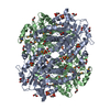
| ||||||||||||
| Unit cell |
| ||||||||||||
| Components on special symmetry positions |
|
-Components
-Protein , 2 types, 2 molecules AB
| #1: Protein | Mass: 43474.820 Da / Num. of mol.: 1 Source method: isolated from a genetically manipulated source Source: (gene. exp.) Rhodococcus wratislaviensis (bacteria) / Gene: Rhow_000804 / Production host: |
|---|---|
| #2: Protein | Mass: 42600.473 Da / Num. of mol.: 1 Source method: isolated from a genetically manipulated source Source: (gene. exp.) Rhodococcus wratislaviensis (bacteria) / Gene: Rhow_000803 / Production host: |
-Non-polymers , 5 types, 149 molecules 


| #3: Chemical | ChemComp-ZN /  Mass: 65.409 Da / Num. of mol.: 1 / Source method: obtained synthetically / Formula: Zn Mass: 65.409 Da / Num. of mol.: 1 / Source method: obtained synthetically / Formula: Zn | ||||||
|---|---|---|---|---|---|---|---|
| #4: Chemical | ChemComp-SO4 /  Mass: 96.063 Da / Num. of mol.: 5 / Source method: obtained synthetically / Formula: SO4 Mass: 96.063 Da / Num. of mol.: 5 / Source method: obtained synthetically / Formula: SO4#5: Chemical | ChemComp-EDO /  Mass: 62.068 Da / Num. of mol.: 17 / Source method: obtained synthetically / Formula: C2H6O2 / Feature type: SUBJECT OF INVESTIGATION Mass: 62.068 Da / Num. of mol.: 17 / Source method: obtained synthetically / Formula: C2H6O2 / Feature type: SUBJECT OF INVESTIGATION#6: Chemical |  Mass: 55.845 Da / Num. of mol.: 2 / Source method: obtained synthetically / Formula: Fe Mass: 55.845 Da / Num. of mol.: 2 / Source method: obtained synthetically / Formula: Fe#7: Water | ChemComp-HOH / | Mass: 18.015 Da / Num. of mol.: 124 / Source method: isolated from a natural source / Formula: H2O |
-Details
| Has ligand of interest | Y |
|---|---|
| Has protein modification | Y |
-Experimental details
-Experiment
| Experiment | Method: X-RAY DIFFRACTION / Number of used crystals: 1 |
|---|
- Sample preparation
| Crystal | Density Matthews: 3.41 Å3/Da / Density % sol: 63.92 % |
|---|---|
| Crystal grow | Temperature: 293 K / Method: vapor diffusion, sitting drop / pH: 8.5 Details: 0.1 M Tris/HCl (pH 8.5), 1 M ammonium sulfate and 12% (v/v) glycerol |
-Data collection
| Diffraction | Mean temperature: 100 K / Serial crystal experiment: N |
|---|---|
| Diffraction source | Source: SYNCHROTRON / Site: SPring-8  / Beamline: BL26B1 / Wavelength: 1 Å / Beamline: BL26B1 / Wavelength: 1 Å |
| Detector | Type: DECTRIS EIGER R 4M / Detector: PIXEL / Date: Jan 31, 2020 |
| Radiation | Protocol: SINGLE WAVELENGTH / Monochromatic (M) / Laue (L): M / Scattering type: x-ray |
| Radiation wavelength | Wavelength: 1 Å / Relative weight: 1 |
| Reflection | Resolution: 2.45→45.92 Å / Num. obs: 207203 / % possible obs: 96.8 % / Redundancy: 2.56 % / Biso Wilson estimate: 40.25 Å2 / CC1/2: 0.994 / Rmerge(I) obs: 0.089 / Rrim(I) all: 0.113 / Net I/σ(I): 9.2 |
| Reflection shell | Resolution: 2.45→2.6 Å / Redundancy: 2.64 % / Rmerge(I) obs: 0.471 / Mean I/σ(I) obs: 1.98 / Num. unique obs: 13330 / CC1/2: 0.709 / Rrim(I) all: 0.593 / % possible all: 98.9 |
- Processing
| Software |
| ||||||||||||||||||||||||||||||||||||||||||||||||||||||||||||||||||||||||||||||||||||||||||||||||||||||||||||||||
|---|---|---|---|---|---|---|---|---|---|---|---|---|---|---|---|---|---|---|---|---|---|---|---|---|---|---|---|---|---|---|---|---|---|---|---|---|---|---|---|---|---|---|---|---|---|---|---|---|---|---|---|---|---|---|---|---|---|---|---|---|---|---|---|---|---|---|---|---|---|---|---|---|---|---|---|---|---|---|---|---|---|---|---|---|---|---|---|---|---|---|---|---|---|---|---|---|---|---|---|---|---|---|---|---|---|---|---|---|---|---|---|---|---|
| Refinement | Method to determine structure: MOLECULAR REPLACEMENT Starting model: 6M1W Resolution: 2.45→44.99 Å / SU ML: 0.2704 / Cross valid method: FREE R-VALUE / σ(F): 1.34 / Phase error: 23.5781
| ||||||||||||||||||||||||||||||||||||||||||||||||||||||||||||||||||||||||||||||||||||||||||||||||||||||||||||||||
| Solvent computation | Shrinkage radii: 0.9 Å / VDW probe radii: 1.11 Å | ||||||||||||||||||||||||||||||||||||||||||||||||||||||||||||||||||||||||||||||||||||||||||||||||||||||||||||||||
| Displacement parameters | Biso mean: 42.46 Å2 | ||||||||||||||||||||||||||||||||||||||||||||||||||||||||||||||||||||||||||||||||||||||||||||||||||||||||||||||||
| Refinement step | Cycle: LAST / Resolution: 2.45→44.99 Å
| ||||||||||||||||||||||||||||||||||||||||||||||||||||||||||||||||||||||||||||||||||||||||||||||||||||||||||||||||
| Refine LS restraints |
| ||||||||||||||||||||||||||||||||||||||||||||||||||||||||||||||||||||||||||||||||||||||||||||||||||||||||||||||||
| LS refinement shell |
|
