 Movie
Movie Controller


+ Open data

- Basic information
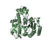
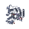
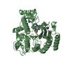
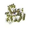
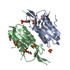
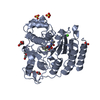
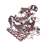
| Entry | Database: PDB / ID: 6lye | |||||||||
|---|---|---|---|---|---|---|---|---|---|---|
| Title | Crystal Structure of mimivirus UNG Y322F in complex with UGI | |||||||||
 Components Components |
| |||||||||
 Keywords Keywords | DNA BINDING PROTEIN/INHIBITOR / UDG / UNG / uracil DNA glycosylase / UGI / DNA BINDING PROTEIN-INHIBITOR complex | |||||||||
| Function / homology |  Function and homology information Function and homology informationbase-excision repair, AP site formation via deaminated base removal / uracil DNA N-glycosylase activity / Hydrolases; Glycosylases; Hydrolysing N-glycosyl compounds Similarity search - Function | |||||||||
| Biological species |  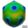 Acanthamoeba polyphaga mimivirusBacillus phage PBS2 (virus) Acanthamoeba polyphaga mimivirusBacillus phage PBS2 (virus) | |||||||||
| Method |  X-RAY DIFFRACTION / SYNCHROTRON / MOLECULAR REPLACEMENT / Resolution: 3.1 Å X-RAY DIFFRACTION / SYNCHROTRON / MOLECULAR REPLACEMENT / Resolution: 3.1 Å | |||||||||
 Authors Authors | Pathak, D. / Kwon, E. / Kim, D.Y. | |||||||||
| Funding support |  Korea, Republic Of, 2items Korea, Republic Of, 2items
| |||||||||
 Citation Citation | Journal: J.Struct.Biol. / Year: 2020 Title: Selective interactions between mimivirus uracil-DNA glycosylase and inhibitory proteins determined by a single amino acid. Authors: Pathak, D. / Kwon, E. / Kim, D.Y. | |||||||||
| History |
|
- Structure visualization
| Structure viewer | Molecule: MolmilJmol/JSmol |
|---|
- Downloads & links
-Download
| PDBx/mmCIF format | 6lye.cif.gz | 84.7 KB | Display | PDBx/mmCIF format |
|---|---|---|---|---|
| PDB format | pdb6lye.ent.gz | 62.3 KB | Display | PDB format |
| PDBx/mmJSON format | 6lye.json.gz | Tree view | PDBx/mmJSON format | |
| Others |  Other downloads Other downloads |
-Validation report
| Arichive directory | https://data.pdbj.org/pub/pdb/validation_reports/ly/6lyeftp://data.pdbj.org/pub/pdb/validation_reports/ly/6lye | HTTPS FTP |
|---|
-Related structure data
| Related structure data | 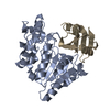 6lydC 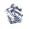 5x55S S: Starting model for refinement C: citing same article ( |
|---|---|
| Similar structure data |


-Links
 PDBj
PDBj- Assembly
| Deposited unit | 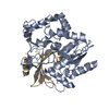
| ||||||||
|---|---|---|---|---|---|---|---|---|---|
| 1 |
| ||||||||
| Unit cell |
|
-Components
| #1: Protein | Mass: 31831.734 Da / Num. of mol.: 1 / Mutation: Y322F Source method: isolated from a genetically manipulated source Source: (gene. exp.) Acanthamoeba polyphaga mimivirus / Gene: UNG, MIMI_L249 / Production host:  References: UniProt: Q5UPT2, Hydrolases; Glycosylases; Hydrolysing N-glycosyl compounds |
|---|---|
| #2: Protein | Mass: 9351.478 Da / Num. of mol.: 1 Source method: isolated from a genetically manipulated source Source: (gene. exp.) Bacillus phage PBS2 (virus) / Gene: UGI / Production host: |
-Experimental details
-Experiment
| Experiment | Method: X-RAY DIFFRACTION / Number of used crystals: 1 |
|---|
- Sample preparation
| Crystal | Density Matthews: 3.16 Å3/Da / Density % sol: 61.14 % |
|---|---|
| Crystal grow | Temperature: 295 K / Method: batch mode Details: 16.75% (v/v) PEG3350, 8% (v/v) PEG400, and 0.1 M sodium acetate/acetic acid pH 5.5 |
-Data collection
| Diffraction | Mean temperature: 80 K / Serial crystal experiment: N |
|---|---|
| Diffraction source | Source: SYNCHROTRON / Site: PAL/PLS / Beamline: 11C / Wavelength: 0.97933 Å |
| Detector | Type: DECTRIS PILATUS3 S 6M / Detector: PIXEL / Date: Mar 12, 2019 |
| Radiation | Protocol: SINGLE WAVELENGTH / Monochromatic (M) / Laue (L): M / Scattering type: x-ray |
| Radiation wavelength | Wavelength: 0.97933 Å / Relative weight: 1 |
| Reflection twin | Operator: h,-h-k,-l / Fraction: 0.34 |
| Reflection | Resolution: 3.1→45.133 Å / Num. obs: 9163 / % possible obs: 97.6 % / Redundancy: 4.25 % / Rmerge(I) obs: 0.127 / Net I/σ(I): 8.3 |
| Reflection shell | Resolution: 3.1→3.31 Å / Rmerge(I) obs: 0.604 / Num. unique obs: 1685 |
- Processing
| Software |
| ||||||||||||||||||||||||||||
|---|---|---|---|---|---|---|---|---|---|---|---|---|---|---|---|---|---|---|---|---|---|---|---|---|---|---|---|---|---|
| Refinement | Method to determine structure: MOLECULAR REPLACEMENT Starting model: 5X55 Resolution: 3.1→45.133 Å / Cross valid method: THROUGHOUT / σ(F): 79.49 / Phase error: 25 / Stereochemistry target values: TWIN_LSQ_F
| ||||||||||||||||||||||||||||
| Solvent computation | Shrinkage radii: 0.9 Å / VDW probe radii: 1.11 Å / Solvent model: FLAT BULK SOLVENT MODEL | ||||||||||||||||||||||||||||
| Displacement parameters | Biso max: 153.15 Å2 / Biso mean: 75.172 Å2 / Biso min: 39.46 Å2 | ||||||||||||||||||||||||||||
| Refinement step | Cycle: final / Resolution: 3.1→45.133 Å
| ||||||||||||||||||||||||||||
| LS refinement shell | Refine-ID: X-RAY DIFFRACTION / Rfactor Rfree error: 0 / Total num. of bins used: 3
|
