- PDB-6lvr: Crystal structure of the PPR domain of Arabidopsis thaliana prote... -
+
Open data
ID or keywords:
Loading...
-
Basic information
Entry
Database: PDB / ID: 6lvr
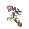
Title
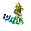
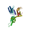
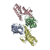
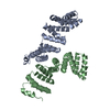
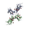
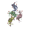
Crystal structure of the PPR domain of Arabidopsis thaliana protein-only RNase P 1 (PRORP1) in complex with tRNA
Components
Proteinaceous RNase P 1, chloroplastic/mitochondrial
yeast phenylalanine tRNA
Keywords
RNA BINDING PROTEIN / Protein-only RNase P tRNA processing tRNA complex Pentatricopeptide repeat convergent evolution
Function / homology
Function and homology information
ribonuclease P / ribonuclease P activity / tRNA 5'-leader removal / tRNA processing / chloroplast / mitochondrion / metal ion binding Similarity search - Function
Protein-only RNase P, C-terminal / Protein-only RNase P / Pentacotripeptide-repeat region of PRORP / Pentacotripeptide-repeat region of PRORP / Pentatricopeptide (PPR) repeat profile. / Pentatricopeptide repeat / Tetratricopeptide repeat domain / Serine Threonine Protein Phosphatase 5, Tetratricopeptide repeat / Alpha Horseshoe / Tetratricopeptide-like helical domain superfamily / Mainly Alpha Similarity search - Domain/homology
In the structure databanks used in Yorodumi, some data are registered as the other names, "COVID-19 virus" and "2019-nCoV". Here are the details of the virus and the list of structure data.
Jan 31, 2019. EMDB accession codes are about to change! (news from PDBe EMDB page)
EMDB accession codes are about to change! (news from PDBe EMDB page)
The allocation of 4 digits for EMDB accession codes will soon come to an end. Whilst these codes will remain in use, new EMDB accession codes will include an additional digit and will expand incrementally as the available range of codes is exhausted. The current 4-digit format prefixed with “EMD-” (i.e. EMD-XXXX) will advance to a 5-digit format (i.e. EMD-XXXXX), and so on. It is currently estimated that the 4-digit codes will be depleted around Spring 2019, at which point the 5-digit format will come into force.
The EM Navigator/Yorodumi systems omit the EMD- prefix.
Related info.:Q: What is EMD? / ID/Accession-code notation in Yorodumi/EM Navigator
Yorodumi is a browser for structure data from EMDB, PDB, SASBDB, etc.
This page is also the successor to EM Navigator detail page, and also detail information page/front-end page for Omokage search.
The word "yorodu" (or yorozu) is an old Japanese word meaning "ten thousand". "mi" (miru) is to see.
Related info.:EMDB / PDB / SASBDB / Comparison of 3 databanks / Yorodumi Search / Aug 31, 2016. New EM Navigator & Yorodumi / Yorodumi Papers / Jmol/JSmol / Function and homology information / Changes in new EM Navigator and Yorodumi
 Movie
Movie Controller

 Yorodumi
 Open data
 Basic information
 Components
 Keywords
 Function and homology information

 X-RAY DIFFRACTION /
  Authors
 United States, 1items
  Citation
 Structure visualization
 Downloads & links
 Other downloads
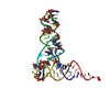


 PDBj
PDBj


 Assembly
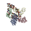


 Mass: 18.015 Da / Num. of mol.: 35 / Source method: isolated from a natural source / Formula: H2O
 Sample preparation
 Processing