 Movie
Movie Controller

[English] 日本語
 Yorodumi
- PDB-6lsc: Structure of the E202Y mutant of the Cl-/H+ antiporter CLC-ec1 fr... -
+ Open data

- Basic information
| Entry | Database: PDB / ID: 6lsc | |||||||||
|---|---|---|---|---|---|---|---|---|---|---|
| Title | Structure of the E202Y mutant of the Cl-/H+ antiporter CLC-ec1 from E.coli: a re-refined model of the 4FTP model | |||||||||
 Components Components | H(+)/Cl(-) exchange transporter ClcA | |||||||||
 Keywords Keywords | MEMBRANE PROTEIN / CLC chloride/proton antiporter | |||||||||
| Function / homology |  Function and homology information Function and homology informationcellular stress response to acidic pH / chloride:proton antiporter activity / voltage-gated chloride channel activity / proton transmembrane transport / chloride transmembrane transport / identical protein binding / plasma membrane Similarity search - Function | |||||||||
| Biological species |  | |||||||||
| Method |  X-RAY DIFFRACTION / SYNCHROTRON / MOLECULAR REPLACEMENT / Resolution: 3.21 Å X-RAY DIFFRACTION / SYNCHROTRON / MOLECULAR REPLACEMENT / Resolution: 3.21 Å | |||||||||
 Authors Authors | Lim, H.H. / Shane, T. / Miller, C. | |||||||||
 Citation Citation | Journal: PLoS Biol. / Year: 2012 Title: Intracellular proton access in a Cl(-)/H(+) antiporter. Authors: Lim, H.H. / Shane, T. / Miller, C. | |||||||||
| History |
|
- Structure visualization
| Structure viewer | Molecule: MolmilJmol/JSmol |
|---|
- Downloads & links
-Download
| PDBx/mmCIF format | 6lsc.cif.gz | 92.3 KB | Display | PDBx/mmCIF format |
|---|---|---|---|---|
| PDB format | pdb6lsc.ent.gz | 69 KB | Display | PDB format |
| PDBx/mmJSON format | 6lsc.json.gz | Tree view | PDBx/mmJSON format | |
| Others |  Other downloads Other downloads |
-Validation report
| Arichive directory | https://data.pdbj.org/pub/pdb/validation_reports/ls/6lscftp://data.pdbj.org/pub/pdb/validation_reports/ls/6lsc | HTTPS FTP |
|---|
-Related structure data
| Related structure data | 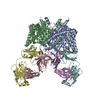 1otsS S: Starting model for refinement |
|---|---|
| Similar structure data |


-Links
 PDBj
PDBj- Assembly
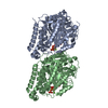
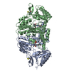
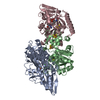
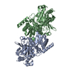
| Deposited unit | 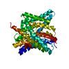
| ||||||||
|---|---|---|---|---|---|---|---|---|---|
| 1 | 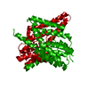
| ||||||||
| Unit cell |
|
-Components
| #1: Protein | Mass: 49692.754 Da / Num. of mol.: 1 / Mutation: E202Y Source method: isolated from a genetically manipulated source Source: (gene. exp.) Strain: K12 / Gene: clcA, eriC, yadQ, b0155, JW5012 / Production host: |
|---|---|
| #2: Chemical | ChemComp-CL /   Mass: 35.453 Da / Num. of mol.: 1 / Source method: obtained synthetically / Formula: Cl / Feature type: SUBJECT OF INVESTIGATION Mass: 35.453 Da / Num. of mol.: 1 / Source method: obtained synthetically / Formula: Cl / Feature type: SUBJECT OF INVESTIGATION |
| #3: Water | ChemComp-HOH /  Mass: 18.015 Da / Num. of mol.: 1 / Source method: isolated from a natural source / Formula: H2O Mass: 18.015 Da / Num. of mol.: 1 / Source method: isolated from a natural source / Formula: H2O |
| Has ligand of interest | Y |
-Experimental details
-Experiment
| Experiment | Method: X-RAY DIFFRACTION / Number of used crystals: 1 |
|---|
- Sample preparation
| Crystal | Density Matthews: 3.8 Å3/Da / Density % sol: 67.6 % |
|---|---|
| Crystal grow | Temperature: 295 K / Method: vapor diffusion, sitting drop / pH: 9.5 Details: 38% PEG400, 100mM CaCl2, 100mM Glycine-NaOH, pH 9.5 |
-Data collection
| Diffraction | Mean temperature: 80 K / Serial crystal experiment: N |
|---|---|
| Diffraction source | Source: SYNCHROTRON / Site: ALS  / Beamline: 5.0.2 / Wavelength: 1 Å / Beamline: 5.0.2 / Wavelength: 1 Å |
| Detector | Type: ADSC QUANTUM 315r / Detector: CCD / Date: Apr 4, 2012 |
| Radiation | Monochromator: Double-crystal, Si(111) / Protocol: SINGLE WAVELENGTH / Monochromatic (M) / Laue (L): M / Scattering type: x-ray |
| Radiation wavelength | Wavelength: 1 Å / Relative weight: 1 |
| Reflection | Resolution: 3.2→25 Å / Num. obs: 12142 / % possible obs: 99 % / Redundancy: 7.3 % / Rmerge(I) obs: 0.16 / Net I/σ(I): 2 |
| Reflection shell | Resolution: 3.2→3.26 Å / Redundancy: 7.2 % / Rmerge(I) obs: 0.59 / Num. unique obs: 904 / % possible all: 99.9 |
- Processing
| Software |
| ||||||||||||||||||||||||||||||||||||||||||||||||||||||||||||
|---|---|---|---|---|---|---|---|---|---|---|---|---|---|---|---|---|---|---|---|---|---|---|---|---|---|---|---|---|---|---|---|---|---|---|---|---|---|---|---|---|---|---|---|---|---|---|---|---|---|---|---|---|---|---|---|---|---|---|---|---|---|
| Refinement | Method to determine structure: MOLECULAR REPLACEMENT Starting model: 1OTS Resolution: 3.21→24.43 Å / Cor.coef. Fo:Fc: 0.922 / Cor.coef. Fo:Fc free: 0.879 / SU B: 18.55 / SU ML: 0.318 / Cross valid method: THROUGHOUT / σ(F): 0 / ESU R Free: 0.451 Details: HYDROGENS HAVE BEEN ADDED IN THE RIDING POSITIONS U VALUES : REFINED INDIVIDUALLY
| ||||||||||||||||||||||||||||||||||||||||||||||||||||||||||||
| Solvent computation | Ion probe radii: 0.8 Å / Shrinkage radii: 0.8 Å / VDW probe radii: 1.2 Å | ||||||||||||||||||||||||||||||||||||||||||||||||||||||||||||
| Displacement parameters | Biso max: 239.86 Å2 / Biso mean: 96.111 Å2 / Biso min: 57.73 Å2
| ||||||||||||||||||||||||||||||||||||||||||||||||||||||||||||
| Refinement step | Cycle: final / Resolution: 3.21→24.43 Å
| ||||||||||||||||||||||||||||||||||||||||||||||||||||||||||||
| Refine LS restraints |
| ||||||||||||||||||||||||||||||||||||||||||||||||||||||||||||
| LS refinement shell | Resolution: 3.213→3.296 Å / Rfactor Rfree error: 0 / Total num. of bins used: 20
|
