 Movie
Movie Controller

[English] 日本語
 Yorodumi
- PDB-6lnw: Crystal structure of accessory secretory protein 1,2 and 3 in Str... -
+ Open data

- Basic information
| Entry | Database: PDB / ID: 6lnw | |||||||||
|---|---|---|---|---|---|---|---|---|---|---|
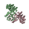
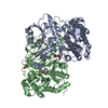
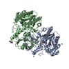
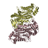
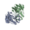
| Title | Crystal structure of accessory secretory protein 1,2 and 3 in Streptococcus pneumoniae | |||||||||
 Components Components |
| |||||||||
 Keywords Keywords | TRANSPORT PROTEIN / Complex | |||||||||
| Function / homology |  Function and homology information Function and homology information | |||||||||
| Biological species |  Streptococcus pneumoniae TIGR4 (bacteria) Streptococcus pneumoniae TIGR4 (bacteria) | |||||||||
| Method |  X-RAY DIFFRACTION / SYNCHROTRON / MOLECULAR REPLACEMENT / Resolution: 2.9 Å X-RAY DIFFRACTION / SYNCHROTRON / MOLECULAR REPLACEMENT / Resolution: 2.9 Å | |||||||||
 Authors Authors | Guo, C. / Feng, Z. / Zuo, G. | |||||||||
| Funding support |  China, 2items China, 2items
| |||||||||
 Citation Citation | Journal: Biochem.Biophys.Res.Commun. / Year: 2020 Title: Structural and functional insights into the Asp1/2/3 complex mediated secretion of pneumococcal serine-rich repeat protein PsrP. Authors: Guo, C. / Feng, Z. / Zuo, G. / Jiang, Y.L. / Zhou, C.Z. / Chen, Y. / Hou, W.T. | |||||||||
| History |
|
- Structure visualization
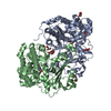
| Structure viewer | Molecule: MolmilJmol/JSmol |
|---|
- Downloads & links
-Download
| PDBx/mmCIF format | 6lnw.cif.gz | 180 KB | Display | PDBx/mmCIF format |
|---|---|---|---|---|
| PDB format | pdb6lnw.ent.gz | 136.3 KB | Display | PDB format |
| PDBx/mmJSON format | 6lnw.json.gz | Tree view | PDBx/mmJSON format | |
| Others |  Other downloads Other downloads |
-Validation report
| Arichive directory | https://data.pdbj.org/pub/pdb/validation_reports/ln/6lnwftp://data.pdbj.org/pub/pdb/validation_reports/ln/6lnw | HTTPS FTP |
|---|
-Related structure data
| Related structure data |  5vaeS S: Starting model for refinement |
|---|---|
| Similar structure data |


-Links
 PDBj
PDBj- Assembly
| Deposited unit | 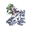
| ||||||||
|---|---|---|---|---|---|---|---|---|---|
| 1 |
| ||||||||
| Unit cell |
|
-Components
| #1: Protein | Mass: 62189.645 Da / Num. of mol.: 1 Source method: isolated from a genetically manipulated source Source: (gene. exp.) Streptococcus pneumoniae TIGR4 (bacteria)Strain: TIGR4 / Gene: SP_1762 / Production host: |
|---|---|
| #2: Protein | Mass: 58828.172 Da / Num. of mol.: 1 Source method: isolated from a genetically manipulated source Source: (gene. exp.) Streptococcus pneumoniae TIGR4 (bacteria)Strain: TIGR4 / Gene: SP_1761 / Production host: |
| #3: Protein | Mass: 18235.680 Da / Num. of mol.: 1 Source method: isolated from a genetically manipulated source Source: (gene. exp.) Streptococcus pneumoniae TIGR4 (bacteria)Strain: TIGR4 / Gene: SP_1760 / Production host: |
| #4: Water | ChemComp-HOH /  Mass: 18.015 Da / Num. of mol.: 90 / Source method: isolated from a natural source / Formula: H2O Mass: 18.015 Da / Num. of mol.: 90 / Source method: isolated from a natural source / Formula: H2O |
-Experimental details
-Experiment
| Experiment | Method: X-RAY DIFFRACTION / Number of used crystals: 1 |
|---|
- Sample preparation
| Crystal | Density Matthews: 2.1 Å3/Da / Density % sol: 41.54 % |
|---|---|
| Crystal grow | Temperature: 293.15 K / Method: vapor diffusion, hanging drop / Details: 0.1 M Tris pH 8.0, 15% PEG 6000 |
-Data collection
| Diffraction | Mean temperature: 100 K / Serial crystal experiment: N |
|---|---|
| Diffraction source | Source: SYNCHROTRON / Site: SSRF / Beamline: BL17U / Wavelength: 0.97915 Å |
| Detector | Type: ADSC QUANTUM 315r / Detector: CCD / Date: Apr 20, 2017 |
| Radiation | Protocol: SINGLE WAVELENGTH / Monochromatic (M) / Laue (L): M / Scattering type: x-ray |
| Radiation wavelength | Wavelength: 0.97915 Å / Relative weight: 1 |
| Reflection | Resolution: 2.9→50 Å / Num. obs: 26531 / % possible obs: 99.85 % / Redundancy: 4.7 % / CC1/2: 0.991 / CC star: 0.998 / Net I/σ(I): 12.25 |
| Reflection shell | Resolution: 2.9→3 Å / Num. unique obs: 2586 / CC1/2: 0.833 / CC star: 0.953 |
- Processing
| Software |
| ||||||||||||||||||||||||||||||||||||||||||||||||||||||||||||
|---|---|---|---|---|---|---|---|---|---|---|---|---|---|---|---|---|---|---|---|---|---|---|---|---|---|---|---|---|---|---|---|---|---|---|---|---|---|---|---|---|---|---|---|---|---|---|---|---|---|---|---|---|---|---|---|---|---|---|---|---|---|
| Refinement | Method to determine structure: MOLECULAR REPLACEMENT Starting model: 5vae Resolution: 2.9→42.94 Å / Cor.coef. Fo:Fc: 0.922 / Cor.coef. Fo:Fc free: 0.873 / WRfactor Rfree: 0.2235 / WRfactor Rwork: 0.187 / FOM work R set: 0.8202 / SU Rfree: 0.3623 / Cross valid method: THROUGHOUT / σ(F): 0 / ESU R Free: 0.362 / Stereochemistry target values: MAXIMUM LIKELIHOOD Details: HYDROGENS HAVE BEEN ADDED IN THE RIDING POSITIONS U VALUES : REFINED INDIVIDUALLY SF FILE CONTAINS FRIEDEL PAIRS UNDER I/F_MINUS AND I/F_PLUS COLUMNS.
| ||||||||||||||||||||||||||||||||||||||||||||||||||||||||||||
| Solvent computation | Ion probe radii: 0.8 Å / Shrinkage radii: 0.8 Å / VDW probe radii: 1.2 Å / Solvent model: MASK | ||||||||||||||||||||||||||||||||||||||||||||||||||||||||||||
| Displacement parameters | Biso max: 165.02 Å2 / Biso mean: 32.37 Å2 / Biso min: 8.15 Å2
| ||||||||||||||||||||||||||||||||||||||||||||||||||||||||||||
| Refinement step | Cycle: final / Resolution: 2.9→42.94 Å
| ||||||||||||||||||||||||||||||||||||||||||||||||||||||||||||
| Refine LS restraints |
| ||||||||||||||||||||||||||||||||||||||||||||||||||||||||||||
| LS refinement shell | Resolution: 2.901→2.976 Å / Rfactor Rfree error: 0 / Total num. of bins used: 20
|
