 Movie
Movie Controller


+ Open data

- Basic information
| Entry | Database: PDB / ID: 6lkk | ||||||
|---|---|---|---|---|---|---|---|
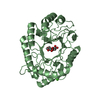
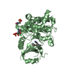
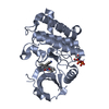
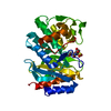
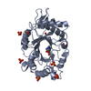
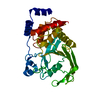
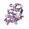
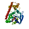
| Title | Two-component system protein mediate signal transduction | ||||||
 Components Components | ABC transporter, solute-binding protein | ||||||
 Keywords Keywords | SIGNALING PROTEIN / two-component system / G6P sensor / signal transduction | ||||||
| Function / homology |  Function and homology information Function and homology informationthiamine binding / thiamine transport / thiamine pyrophosphate binding / outer membrane-bounded periplasmic space Similarity search - Function | ||||||
| Biological species |   Staphylococcus aureus (bacteria) Staphylococcus aureus (bacteria) | ||||||
| Method |  X-RAY DIFFRACTION / SYNCHROTRON / SAD / Resolution: 1.502 Å X-RAY DIFFRACTION / SYNCHROTRON / SAD / Resolution: 1.502 Å | ||||||
 Authors Authors | Wang, M. / Tao, Y. | ||||||
 Citation Citation | Journal: Proc.Natl.Acad.Sci.USA / Year: 2020 Title: Interface switch mediates signal transmission in a two-component system. Authors: Wang, M. / Guo, Q. / Zhu, K. / Fang, B. / Yang, Y. / Teng, M. / Li, X. / Tao, Y. | ||||||
| History |
|
- Structure visualization
| Structure viewer | Molecule: MolmilJmol/JSmol |
|---|
- Downloads & links
-Download
| PDBx/mmCIF format | 6lkk.cif.gz | 132 KB | Display | PDBx/mmCIF format |
|---|---|---|---|---|
| PDB format | pdb6lkk.ent.gz | 102.9 KB | Display | PDB format |
| PDBx/mmJSON format | 6lkk.json.gz | Tree view | PDBx/mmJSON format | |
| Others |  Other downloads Other downloads |
-Validation report
| Arichive directory | https://data.pdbj.org/pub/pdb/validation_reports/lk/6lkkftp://data.pdbj.org/pub/pdb/validation_reports/lk/6lkk | HTTPS FTP |
|---|
-Related structure data


-Links
 PDBj
PDBj


- Assembly
| Deposited unit | 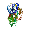
| ||||||||
|---|---|---|---|---|---|---|---|---|---|
| 1 |
| ||||||||
| Unit cell |
|
-Components
| #1: Protein | Mass: 33754.316 Da / Num. of mol.: 1 Source method: isolated from a genetically manipulated source Source: (gene. exp.) Staphylococcus aureus (bacteria)Gene: E4U00_07700, EP54_06205, EQ90_12025, HMPREF3211_02751, NCTC10654_00249, NCTC10702_00414, RK64_01575 Production host: |
|---|---|
| #2: Sugar | ChemComp-G6P /   Type: D-saccharide, alpha linking / Mass: 260.136 Da / Num. of mol.: 1 Type: D-saccharide, alpha linking / Mass: 260.136 Da / Num. of mol.: 1Source method: isolated from a genetically manipulated source Formula: C6H13O9P / Feature type: SUBJECT OF INVESTIGATION |
| #3: Water | ChemComp-HOH /  Mass: 18.015 Da / Num. of mol.: 315 / Source method: isolated from a natural source / Formula: H2O Mass: 18.015 Da / Num. of mol.: 315 / Source method: isolated from a natural source / Formula: H2O |
| Has ligand of interest | Y |
-Experimental details
-Experiment
| Experiment | Method: X-RAY DIFFRACTION / Number of used crystals: 1 |
|---|
- Sample preparation
| Crystal | Density Matthews: 2.28 Å3/Da / Density % sol: 45.97 % |
|---|---|
| Crystal grow | Temperature: 293 K / Method: vapor diffusion / Details: 0.1M Tris pH 7.5-7.9, 21% PEG 3350 / PH range: 7.5-7.9 |
-Data collection
| Diffraction | Mean temperature: 100 K / Serial crystal experiment: N |
|---|---|
| Diffraction source | Source: SYNCHROTRON / Site: SSRF  / Beamline: BL19U1 / Wavelength: 0.9793 Å / Beamline: BL19U1 / Wavelength: 0.9793 Å |
| Detector | Type: DECTRIS PILATUS3 6M / Detector: PIXEL / Date: Sep 5, 2016 |
| Radiation | Protocol: SINGLE WAVELENGTH / Monochromatic (M) / Laue (L): M / Scattering type: x-ray |
| Radiation wavelength | Wavelength: 0.9793 Å / Relative weight: 1 |
| Reflection | Resolution: 1.5→50 Å / Num. obs: 47810 / % possible obs: 99.9 % / Redundancy: 7.1 % / CC1/2: 0.997 / Rmerge(I) obs: 0.065 / Rpim(I) all: 0.021 / Rrim(I) all: 0.074 / Net I/σ(I): 15.36 |
| Reflection shell | Resolution: 1.5→1.56 Å / Redundancy: 6.7 % / Rmerge(I) obs: 0.451 / Mean I/σ(I) obs: 2.42 / Num. unique obs: 3920 / CC1/2: 0.986 / Rpim(I) all: 0.135 / Rrim(I) all: 0.497 / % possible all: 100 |
- Processing
| Software |
| ||||||||||||||||||||||||||||||||||||||||||||||||||||||||||||||||||||||||||||||||||||||||||||||||||||||||||||
|---|---|---|---|---|---|---|---|---|---|---|---|---|---|---|---|---|---|---|---|---|---|---|---|---|---|---|---|---|---|---|---|---|---|---|---|---|---|---|---|---|---|---|---|---|---|---|---|---|---|---|---|---|---|---|---|---|---|---|---|---|---|---|---|---|---|---|---|---|---|---|---|---|---|---|---|---|---|---|---|---|---|---|---|---|---|---|---|---|---|---|---|---|---|---|---|---|---|---|---|---|---|---|---|---|---|---|---|---|---|
| Refinement | Method to determine structure: SAD / Resolution: 1.502→37.307 Å / SU ML: 0.16 / Cross valid method: THROUGHOUT / σ(F): 1.36 / Phase error: 21.01
| ||||||||||||||||||||||||||||||||||||||||||||||||||||||||||||||||||||||||||||||||||||||||||||||||||||||||||||
| Solvent computation | Shrinkage radii: 0.9 Å / VDW probe radii: 1.11 Å | ||||||||||||||||||||||||||||||||||||||||||||||||||||||||||||||||||||||||||||||||||||||||||||||||||||||||||||
| Displacement parameters | Biso max: 84.91 Å2 / Biso mean: 28.3628 Å2 / Biso min: 13.99 Å2 | ||||||||||||||||||||||||||||||||||||||||||||||||||||||||||||||||||||||||||||||||||||||||||||||||||||||||||||
| Refinement step | Cycle: final / Resolution: 1.502→37.307 Å
| ||||||||||||||||||||||||||||||||||||||||||||||||||||||||||||||||||||||||||||||||||||||||||||||||||||||||||||
| Refine LS restraints |
| ||||||||||||||||||||||||||||||||||||||||||||||||||||||||||||||||||||||||||||||||||||||||||||||||||||||||||||
| LS refinement shell | Refine-ID: X-RAY DIFFRACTION / Rfactor Rfree error: 0
|
