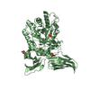
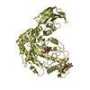
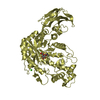
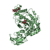
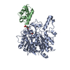
- PDB-6li2: Crystal structure of GPR52 ligand free form with rubredoxin fusion -
+
Open data
ID or keywords:
Loading...
-
Basic information
Entry
Database: PDB / ID: 6li2
Title
Crystal structure of GPR52 ligand free form with rubredoxin fusion
Components
Chimera of G-protein coupled receptor 52 and Rubredoxin
Keywords
MEMBRANE PROTEIN / Human GPR52 receptor / Class A / orphan GPCR / apo form / rubredoxin / LCP
Function / homology
Function and homology information
alkane catabolic process / G protein-coupled photoreceptor activity / cellular response to light stimulus / phototransduction / locomotory behavior / G protein-coupled receptor activity / electron transfer activity / iron ion binding / response to xenobiotic stimulus / G protein-coupled receptor signaling pathway / plasma membrane Similarity search - Function
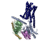
Journal: Nature / Year: 2020 Title: Structural basis of ligand recognition and self-activation of orphan GPR52. Authors: Xi Lin / Mingyue Li / Niandong Wang / Yiran Wu / Zhipu Luo / Shimeng Guo / Gye-Won Han / Shaobai Li / Yang Yue / Xiaohu Wei / Xin Xie / Yong Chen / Suwen Zhao / Jian Wu / Ming Lei / Fei Xu / Abstract: GPR52 is a class-A orphan G-protein-coupled receptor that is highly expressed in the brain and represents a promising therapeutic target for the treatment of Huntington's disease and several ...GPR52 is a class-A orphan G-protein-coupled receptor that is highly expressed in the brain and represents a promising therapeutic target for the treatment of Huntington's disease and several psychiatric disorders. Pathological malfunction of GPR52 signalling occurs primarily through the heterotrimeric G protein, but it is unclear how GPR52 and G couple for signal transduction and whether a native ligand or other activating input is required. Here we present the high-resolution structures of human GPR52 in three states: a ligand-free state, a G-coupled self-activation state and a potential allosteric ligand-bound state. Together, our structures reveal that extracellular loop 2 occupies the orthosteric binding pocket and operates as a built-in agonist, conferring an intrinsically high level of basal activity to GPR52. A fully active state is achieved when G is coupled to GPR52 in the absence of an external agonist. The receptor also features a side pocket for ligand binding. These insights into the structure and function of GPR52 could improve our understanding of other self-activated GPCRs, enable the identification of endogenous and tool ligands, and guide drug discovery efforts that target GPR52.
Method to determine structure: MOLECULAR REPLACEMENT Starting model: Rosetta modelling Resolution: 2.8→29.25 Å / Cor.coef. Fo:Fc: 0.889 / Cor.coef. Fo:Fc free: 0.892 / SU B: 32.117 / SU ML: 0.315 / Cross valid method: THROUGHOUT / ESU R: 16.343 / ESU R Free: 0.401 / Stereochemistry target values: MAXIMUM LIKELIHOOD Details: HYDROGENS HAVE BEEN ADDED IN THE RIDING POSITIONS Authors aniso-corrected the observed data by STARANISO which remove weak reflections.
Rfactor
Num. reflection
% reflection
Selection details
Rfree
0.26295
612
4.9 %
RANDOM
Rwork
0.24112
-
-
-
obs
0.24217
11830
81.1 %
-
Solvent computation
Ion probe radii: 0.8 Å / Shrinkage radii: 0.8 Å / VDW probe radii: 1.2 Å / Solvent model: BABINET MODEL WITH MASK
 Movie
Movie Controller

 Yorodumi
 Open data
 Basic information
 Components
 Keywords
 Function and homology information
 Homo sapiens (human)
 Clostridium pasteurianum (bacteria)
 X-RAY DIFFRACTION /
  Authors
 China, 1items
  Citation
 Structure visualization
 Downloads & links
 Other downloads
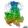


 PDBj
PDBj


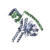
 Assembly

 Spodoptera frugiperda (fall armyworm) / References: UniProt: Q9Y2T5, UniProt: P00268
 Mass: 65.409 Da / Num. of mol.: 1 / Source method: obtained synthetically / Formula: Zn
 Mass: 356.540 Da / Num. of mol.: 11 / Source method: obtained synthetically / Formula: C21H40O4
 Mass: 106.120 Da / Num. of mol.: 1 / Source method: obtained synthetically / Formula: C4H10O3
 Mass: 18.015 Da / Num. of mol.: 18 / Source method: isolated from a natural source / Formula: H2O
 Sample preparation
 / Beamline: BL41XU / Wavelength: 1 Å
 Processing