[English] 日本語
Yorodumi
- PDB-6lfl: Crystal structure of a class A GPCR -

+
Open data


ID or keywords:

Loading...

-
Basic information

Entry
Database: PDB / ID: 6lfl
TitleCrystal structure of a class A GPCR
ComponentsC-X-C chemokine receptor type 2,GlgA glycogen synthase,C-X-C chemokine receptor type 2
KeywordsMEMBRANE PROTEIN / G protein-coupled receptor
Function / homology
Function and homology information


interleukin-8-mediated signaling pathway / metanephric tubule morphogenesis / negative regulation of neutrophil apoptotic process / interleukin-8 receptor activity / mast cell granule / interleukin-8 binding / C-X-C chemokine receptor activity / acute inflammatory response to antigenic stimulus / glycogen (starch) synthase activity / C-C chemokine receptor activity ...interleukin-8-mediated signaling pathway / metanephric tubule morphogenesis / negative regulation of neutrophil apoptotic process / interleukin-8 receptor activity / mast cell granule / interleukin-8 binding / C-X-C chemokine receptor activity / acute inflammatory response to antigenic stimulus / glycogen (starch) synthase activity / C-C chemokine receptor activity / neutrophil activation / C-C chemokine binding / positive regulation of vascular permeability / positive regulation of neutrophil chemotaxis / Chemokine receptors bind chemokines / dendritic cell chemotaxis / midbrain development / positive regulation of cardiac muscle cell apoptotic process / cellular defense response / neutrophil chemotaxis / secretory granule membrane / G protein-coupled receptor activity / calcium-mediated signaling / receptor internalization / mitotic spindle / positive regulation of angiogenesis / microtubule cytoskeleton / chemotaxis / phospholipase C-activating G protein-coupled receptor signaling pathway / G alpha (i) signalling events / positive regulation of cytosolic calcium ion concentration / cell surface receptor signaling pathway / immune response / inflammatory response / external side of plasma membrane / nucleotide binding / positive regulation of cell population proliferation / Neutrophil degranulation / cell surface / signal transduction / nucleoplasm / membrane / plasma membrane
Similarity search - Function
CXC chemokine receptor 2 / CXC chemokine receptor 1/2 / Glycosyl transferases group 1 / Bacterial/plant glycogen synthase / Starch synthase, catalytic domain / Starch synthase catalytic domain / Glycosyl transferases group 1 / G-protein coupled receptors family 1 signature. / G protein-coupled receptor, rhodopsin-like / GPCR, rhodopsin-like, 7TM ...CXC chemokine receptor 2 / CXC chemokine receptor 1/2 / Glycosyl transferases group 1 / Bacterial/plant glycogen synthase / Starch synthase, catalytic domain / Starch synthase catalytic domain / Glycosyl transferases group 1 / G-protein coupled receptors family 1 signature. / G protein-coupled receptor, rhodopsin-like / GPCR, rhodopsin-like, 7TM / G-protein coupled receptors family 1 profile. / 7 transmembrane receptor (rhodopsin family)
Similarity search - Domain/homology
Chem-EBX / C-X-C chemokine receptor type 2 / Glycogen synthase
Similarity search - Component
Biological speciesHomo sapiens (human)
Pyrococcus abyssi (archaea)
MethodX-RAY DIFFRACTION / SYNCHROTRON / MOLECULAR REPLACEMENT / molecular replacement / Resolution: 3.2 Å
AuthorsLiu, Z.J. / Hua, T. / Liu, K.W. / Wu, L.J.
Citation
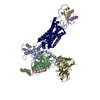
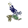
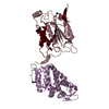
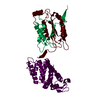
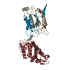
Journal: Nature / Year: 2020
Title: Structural basis of CXC chemokine receptor 2 activation and signalling.
Authors: Kaiwen Liu / Lijie Wu / Shuguang Yuan / Meng Wu / Yueming Xu / Qianqian Sun / Shu Li / Suwen Zhao / Tian Hua / Zhi-Jie Liu /
Abstract: Chemokines and their receptors mediate cell migration, which influences multiple fundamental biological processes and disease conditions such as inflammation and cancer. Although ample effort has ...Chemokines and their receptors mediate cell migration, which influences multiple fundamental biological processes and disease conditions such as inflammation and cancer. Although ample effort has been invested into the structural investigation of the chemokine receptors and receptor-chemokine recognition, less is known about endogenous chemokine-induced receptor activation and G-protein coupling. Here we present the cryo-electron microscopy structures of interleukin-8 (IL-8, also known as CXCL8)-activated human CXC chemokine receptor 2 (CXCR2) in complex with G protein, along with a crystal structure of CXCR2 bound to a designed allosteric antagonist. Our results reveal a unique shallow mode of binding between CXCL8 and CXCR2, and also show the interactions between CXCR2 and G protein. Further structural analysis of the inactive and active states of CXCR2 reveals a distinct activation process and the competitive small-molecule antagonism of chemokine receptors. In addition, our results provide insights into how a G-protein-coupled receptor is activated by an endogenous protein molecule, which will assist in the rational development of therapeutics that target the chemokine system for better pharmacological profiles.
History
DepositionDec 3, 2019Deposition site: PDBJ / Processing site: PDBJ
Revision 1.0Sep 2, 2020Provider: repository / Type: Initial release
Revision 1.1Sep 16, 2020Group: Database references / Category: citation / citation_author
Item: _citation.journal_volume / _citation.page_first ..._citation.journal_volume / _citation.page_first / _citation.page_last / _citation_author.identifier_ORCID
Revision 1.2Nov 22, 2023Group: Data collection / Database references / Refinement description
Category: chem_comp_atom / chem_comp_bond ...chem_comp_atom / chem_comp_bond / database_2 / pdbx_initial_refinement_model
Item: _database_2.pdbx_DOI / _database_2.pdbx_database_accession

-
Structure visualization

Structure viewerMolecule:
MolmilJmol/JSmol

Downloads & links

-
Assembly

Deposited unit
A: C-X-C chemokine receptor type 2,GlgA glycogen synthase,C-X-C chemokine receptor type 2
hetero molecules


Theoretical massNumber of molelcules
Total (without water)57,1212
Polymers56,6951
Non-polymers4261
Water0
1


  • Idetical with deposited unit
  • defined by author
TypeNameSymmetry operationNumber
identity operation1_555x,y,z1
Unit cell
Length a, b, c (Å)41.300, 108.460, 82.600
Angle α, β, γ (deg.)90.000, 91.160, 90.000
Int Tables number4
Space group name H-MP1211

-
Components

#1: Protein C-X-C chemokine receptor type 2,GlgA glycogen synthase,C-X-C chemokine receptor type 2 / CXCR-2 / CDw128b / GRO/MGSA receptor / High affinity interleukin-8 receptor B / IL-8R B / IL-8 ...CXCR-2 / CDw128b / GRO/MGSA receptor / High affinity interleukin-8 receptor B / IL-8R B / IL-8 receptor type 2 / Glycogen synthase / CXCR-2 / CDw128b / GRO/MGSA receptor / High affinity interleukin-8 receptor B / IL-8R B / IL-8 receptor type 2


Mass: 56694.871 Da / Num. of mol.: 1 / Mutation: L135W, A249E, G303A
Source method: isolated from a genetically manipulated source
Source: (gene. exp.) Homo sapiens (human), (gene. exp.) Pyrococcus abyssi (strain GE5 / Orsay) (archaea)
Gene: CXCR2, IL8RB, PAB2292 / Strain: GE5 / Orsay / Production host: 
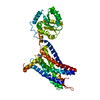
Spodoptera frugiperda (fall armyworm) / References: UniProt: P25025, UniProt: Q9V2J8
#2: Chemical ChemComp-EBX / 4-[[3,4-bis(oxidanylidene)-2-[[(1~{R})-1-(4-propan-2-ylfuran-2-yl)propyl]amino]cyclobuten-1-yl]amino]-~{N},~{N}-dimethyl-3-oxidanyl-pyridine-2-carboxamide


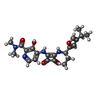
Mass: 426.466 Da / Num. of mol.: 1 / Source method: obtained synthetically / Formula: C22H26N4O5 / Feature type: SUBJECT OF INVESTIGATION
Has ligand of interestY

-
Experimental details

-
Experiment

ExperimentMethod: X-RAY DIFFRACTION / Number of used crystals: 1

-
Sample preparation

CrystalDensity Matthews: 3.26 Å3/Da / Density % sol: 62.3 %
Crystal growTemperature: 293 K / Method: lipidic cubic phase
Details: 100mM HEPES pH7.0, 32% PEG 400, 50-150 mM Sodium tartrate dibasic dihydrate salt

-
Data collection

DiffractionMean temperature: 100 K / Serial crystal experiment: N
Diffraction sourceSource: SYNCHROTRON / Site: SPring-8 / Beamline: BL41XU / Wavelength: 1 Å
DetectorType: DECTRIS EIGER X 16M / Detector: PIXEL / Date: Oct 19, 2018
RadiationProtocol: SINGLE WAVELENGTH / Monochromatic (M) / Laue (L): M / Scattering type: x-ray
Radiation wavelengthWavelength: 1 Å / Relative weight: 1
ReflectionResolution: 3.2→45.33 Å / Num. obs: 12047 / % possible obs: 99.8 % / Redundancy: 10.78 % / Biso Wilson estimate: 110.76 Å2 / CC1/2: 0.996 / Net I/σ(I): 10.06
Reflection shell

Diffraction-ID: 1

Resolution (Å)Redundancy (%)Rmerge(I) obsMean I/σ(I) obsNum. measured obsNum. possibleNum. unique obsCC1/2Rrim(I) all% possible all
3.2-3.397.0340.5991.413252189418840.5450.64499.5
3.39-3.637.9990.4482.3114767185518460.8560.47699.5
3.63-3.929.8570.3274.3916806170617050.9540.34399.9
3.92-4.2911.3610.2477.8817734156315610.9830.25799.9
4.29-4.812.3230.19212.6717548142514240.9870.19999.9
4.8-5.5413.1650.15415.5116549125712570.9950.159100
5.54-6.7914.0970.14817.1815281108410840.9930.153100
6.79-9.613.8410.09126.57113228188180.9980.094100
9.6-45.3314.2240.07835.4366574714680.9960.08199.4

-
Phasing

PhasingMethod: molecular replacement

-
Processing

Software
NameVersionClassificationNB
BUSTER2.10.3refinement
XDSdata reduction
XSCALEdata scaling
PHASERphasing
PDB_EXTRACT3.25data extraction
RefinementMethod to determine structure: MOLECULAR REPLACEMENT
Starting model: 5LWE
Resolution: 3.2→45.33 Å / Cor.coef. Fo:Fc: 0.902 / Cor.coef. Fo:Fc free: 0.926 / Cross valid method: THROUGHOUT / σ(F): 0 / SU Rfree Blow DPI: 0.449
RfactorNum. reflection% reflectionSelection details
Rfree0.264 616 5.11 %RANDOM
Rwork0.231 ---
obs0.233 12047 99.8 %-
Displacement parametersBiso max: 241.9 Å2 / Biso mean: 132.56 Å2 / Biso min: 30 Å2
Baniso -1Baniso -2Baniso -3
1--0.5433 Å20 Å2-5.5759 Å2
2---15.8165 Å20 Å2
3---16.3598 Å2
Refine analyzeLuzzati coordinate error obs: 0.52 Å
Refinement stepCycle: final / Resolution: 3.2→45.33 Å
ProteinNucleic acidLigandSolventTotal
Num. atoms3706 0 31 0 3737
Biso mean--98.56 --
Num. residues----471
Refine LS restraints
Refine-IDTypeNumberRestraint functionWeightDev ideal
X-RAY DIFFRACTIONt_dihedral_angle_d1316SINUSOIDAL2
X-RAY DIFFRACTIONt_trig_c_planes
X-RAY DIFFRACTIONt_gen_planes613HARMONIC5
X-RAY DIFFRACTIONt_it3823HARMONIC20
X-RAY DIFFRACTIONt_nbd
X-RAY DIFFRACTIONt_improper_torsion
X-RAY DIFFRACTIONt_pseud_angle
X-RAY DIFFRACTIONt_chiral_improper_torsion497SEMIHARMONIC5
X-RAY DIFFRACTIONt_sum_occupancies
X-RAY DIFFRACTIONt_utility_distance
X-RAY DIFFRACTIONt_utility_angle
X-RAY DIFFRACTIONt_utility_torsion
X-RAY DIFFRACTIONt_ideal_dist_contact4388SEMIHARMONIC4
X-RAY DIFFRACTIONt_bond_d3823HARMONIC20.008
X-RAY DIFFRACTIONt_angle_deg5173HARMONIC21
X-RAY DIFFRACTIONt_omega_torsion1.93
X-RAY DIFFRACTIONt_other_torsion23.04
LS refinement shellResolution: 3.2→3.5 Å / Rfactor Rfree error: 0 / Total num. of bins used: 6
RfactorNum. reflection% reflection
Rfree0.2562 147 5.14 %
Rwork0.2123 2711 -
all0.2145 2858 -
obs--99.44 %
Refinement TLS params.Method: refined / Origin x: 40.412 Å / Origin y: -19.015 Å / Origin z: 183.59 Å
111213212223313233
T-0.1988 Å20.0337 Å2-0.071 Å2-0.0881 Å20.0018 Å2---0.4302 Å2
L1.0383 °2-1.094 °2-1.2712 °2-2.1584 °21.5478 °2--0.9676 °2
S0.0765 Å °0.2724 Å °-0.0327 Å °-0.1226 Å °-0.1571 Å °-0.0138 Å °-0.3108 Å °-0.1083 Å °0.0807 Å °
Refinement TLS groupSelection details: { A|* }

+
About Yorodumi

-
News

-
Feb 9, 2022. New format data for meta-information of EMDB entries

New format data for meta-information of EMDB entries

  • Version 3 of the EMDB header file is now the official format.
  • The previous official version 1.9 will be removed from the archive.

Related info.:EMDB header

External links:wwPDB to switch to version 3 of the EMDB data model

-
Aug 12, 2020. Covid-19 info

Covid-19 info

URL: https://pdbj.org/emnavi/covid19.php

New page: Covid-19 featured information page in EM Navigator.

Related info.:Covid-19 info / Mar 5, 2020. Novel coronavirus structure data

+
Mar 5, 2020. Novel coronavirus structure data

Novel coronavirus structure data

Related info.:Yorodumi Speices / Aug 12, 2020. Covid-19 info

External links:COVID-19 featured content - PDBj / Molecule of the Month (242):Coronavirus Proteases

+
Jan 31, 2019. EMDB accession codes are about to change! (news from PDBe EMDB page)

EMDB accession codes are about to change! (news from PDBe EMDB page)

  • The allocation of 4 digits for EMDB accession codes will soon come to an end. Whilst these codes will remain in use, new EMDB accession codes will include an additional digit and will expand incrementally as the available range of codes is exhausted. The current 4-digit format prefixed with “EMD-” (i.e. EMD-XXXX) will advance to a 5-digit format (i.e. EMD-XXXXX), and so on. It is currently estimated that the 4-digit codes will be depleted around Spring 2019, at which point the 5-digit format will come into force.
  • The EM Navigator/Yorodumi systems omit the EMD- prefix.

Related info.:Q: What is EMD? / ID/Accession-code notation in Yorodumi/EM Navigator

External links:EMDB Accession Codes are Changing Soon! / Contact to PDBj

+
Jul 12, 2017. Major update of PDB

Major update of PDB

  • wwPDB released updated PDB data conforming to the new PDBx/mmCIF dictionary.
  • This is a major update changing the version number from 4 to 5, and with Remediation, in which all the entries are updated.
  • In this update, many items about electron microscopy experimental information are reorganized (e.g. em_software).
  • Now, EM Navigator and Yorodumi are based on the updated data.

External links:wwPDB Remediation / Enriched Model Files Conforming to OneDep Data Standards Now Available in the PDB FTP Archive

-
Yorodumi

Thousand views of thousand structures

  • Yorodumi is a browser for structure data from EMDB, PDB, SASBDB, etc.
  • This page is also the successor to EM Navigator detail page, and also detail information page/front-end page for Omokage search.
  • The word "yorodu" (or yorozu) is an old Japanese word meaning "ten thousand". "mi" (miru) is to see.

Related info.:EMDB / PDB / SASBDB / Comparison of 3 databanks / Yorodumi Search / Aug 31, 2016. New EM Navigator & Yorodumi / Yorodumi Papers / Jmol/JSmol / Function and homology information / Changes in new EM Navigator and Yorodumi

Read more