 Movie
Movie Controller

[English] 日本語
 Yorodumi
- PDB-6lf9: Crystal structure of pSLA-1*1301 complex with dodecapeptide RVEDV... -
+ Open data

- Basic information
| Entry | Database: PDB / ID: 6lf9 | ||||||
|---|---|---|---|---|---|---|---|
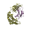
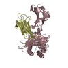
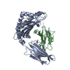
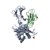
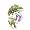
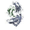
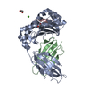
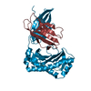
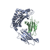
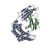
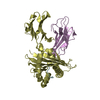
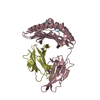
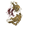
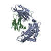
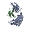
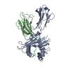
| Title | Crystal structure of pSLA-1*1301 complex with dodecapeptide RVEDVTNTAEYW | ||||||
 Components Components |
| ||||||
 Keywords Keywords | STRUCTURAL PROTEIN / MHC class I structure / A single-amino acid mutation / Peptide motifs / Random peptide library | ||||||
| Function / homology |  Function and homology information Function and homology informationER-Phagosome pathway / Endosomal/Vacuolar pathway / Immunoregulatory interactions between a Lymphoid and a non-Lymphoid cell / DAP12 interactions / DAP12 signaling / Antigen Presentation: Folding, assembly and peptide loading of class I MHC / Neutrophil degranulation / response to metal ion / antigen processing and presentation of peptide antigen via MHC class I / lumenal side of endoplasmic reticulum membrane ...ER-Phagosome pathway / Endosomal/Vacuolar pathway / Immunoregulatory interactions between a Lymphoid and a non-Lymphoid cell / DAP12 interactions / DAP12 signaling / Antigen Presentation: Folding, assembly and peptide loading of class I MHC / Neutrophil degranulation / response to metal ion / antigen processing and presentation of peptide antigen via MHC class I / lumenal side of endoplasmic reticulum membrane / MHC class I protein complex / peptide antigen assembly with MHC class II protein complex / MHC class II protein complex / antigen processing and presentation of exogenous peptide antigen via MHC class II / positive regulation of immune response / peptide antigen binding / phagocytic vesicle membrane / positive regulation of T cell activation / MHC class II protein complex binding / late endosome membrane / immune response / lysosomal membrane / extracellular region Similarity search - Function | ||||||
| Biological species |  synthetic construct (others) | ||||||
| Method |  X-RAY DIFFRACTION / SYNCHROTRON / MOLECULAR REPLACEMENT / Resolution: 2.5 Å X-RAY DIFFRACTION / SYNCHROTRON / MOLECULAR REPLACEMENT / Resolution: 2.5 Å | ||||||
 Authors Authors | Wei, X.H. / Wang, S. / Zhang, N.Z. / Xia, C. | ||||||
 Citation Citation | Journal: Front Immunol / Year: 2022 Title: Peptidomes and Structures Illustrate How SLA-I Micropolymorphism Influences the Preference of Binding Peptide Length. Authors: Wei, X.H. / Wang, S. / Zhang, N.Z. / Xia, C. | ||||||
| History |
|
- Structure visualization
| Structure viewer | Molecule: MolmilJmol/JSmol |
|---|
- Downloads & links
-Download
| PDBx/mmCIF format | 6lf9.cif.gz | 317.2 KB | Display | PDBx/mmCIF format |
|---|---|---|---|---|
| PDB format | pdb6lf9.ent.gz | 259.2 KB | Display | PDB format |
| PDBx/mmJSON format | 6lf9.json.gz | Tree view | PDBx/mmJSON format | |
| Others |  Other downloads Other downloads |
-Validation report
| Arichive directory | https://data.pdbj.org/pub/pdb/validation_reports/lf/6lf9ftp://data.pdbj.org/pub/pdb/validation_reports/lf/6lf9 | HTTPS FTP |
|---|
-Related structure data
| Related structure data |  3qq3S S: Starting model for refinement |
|---|---|
| Similar structure data |


-Links
 PDBj
PDBj

- Assembly
| Deposited unit | 
| ||||||||
|---|---|---|---|---|---|---|---|---|---|
| 1 | 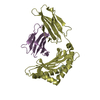
| ||||||||
| 2 | 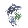
| ||||||||
| 3 | 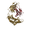
| ||||||||
| 4 | 
| ||||||||
| Unit cell |
|
-Components
| #1: Protein | Mass: 31305.666 Da / Num. of mol.: 4 Source method: isolated from a genetically manipulated source Source: (gene. exp.)  #2: Protein | Mass: 11332.788 Da / Num. of mol.: 4 Source method: isolated from a genetically manipulated source Source: (gene. exp.) #3: Protein/peptide | Mass: 1483.558 Da / Num. of mol.: 4 / Source method: obtained synthetically / Source: (synth.) synthetic construct (others) #4: Water | ChemComp-HOH / |  Mass: 18.015 Da / Num. of mol.: 295 / Source method: isolated from a natural source / Formula: H2O Mass: 18.015 Da / Num. of mol.: 295 / Source method: isolated from a natural source / Formula: H2OHas protein modification | Y | |
|---|
-Experimental details
-Experiment
| Experiment | Method: X-RAY DIFFRACTION / Number of used crystals: 1 |
|---|
- Sample preparation
| Crystal | Density Matthews: 2.38 Å3/Da / Density % sol: 48.35 % |
|---|---|
| Crystal grow | Temperature: 277 K / Method: vapor diffusion, sitting drop / pH: 8 Details: 0.2 M Ammonium fluoride, 20% w/v Polyethylene glycol 3,350 |
-Data collection
| Diffraction | Mean temperature: 100 K / Serial crystal experiment: N |
|---|---|
| Diffraction source | Source: SYNCHROTRON / Site: ALS  / Beamline: 8.3.1 / Wavelength: 0.97931 Å / Beamline: 8.3.1 / Wavelength: 0.97931 Å |
| Detector | Type: ADSC QUANTUM 315r / Detector: CCD / Date: Sep 23, 2017 |
| Radiation | Protocol: SINGLE WAVELENGTH / Monochromatic (M) / Laue (L): M / Scattering type: x-ray |
| Radiation wavelength | Wavelength: 0.97931 Å / Relative weight: 1 |
| Reflection | Resolution: 2.16→199.63 Å / Num. obs: 55584 / % possible obs: 97.1 % / Redundancy: 5.5 % / Rmerge(I) obs: 0.176 / Rsym value: 0.176 / Net I/σ(I): 6 |
| Reflection shell | Resolution: 2.16→2.28 Å / Redundancy: 5.1 % / Rmerge(I) obs: 0.426 / Num. unique obs: 43280 / Rsym value: 0.426 / % possible all: 97.1 |
- Processing
| Software |
| ||||||||||||||||||||
|---|---|---|---|---|---|---|---|---|---|---|---|---|---|---|---|---|---|---|---|---|---|
| Refinement | Method to determine structure: MOLECULAR REPLACEMENT Starting model: 3qq3 Resolution: 2.5→199.63 Å / Cor.coef. Fo:Fc: 0.895 / Cor.coef. Fo:Fc free: 0.86 / SU B: 0.003 / SU ML: 0 / Cross valid method: THROUGHOUT / ESU R: 0.306 / ESU R Free: 0.355 / Stereochemistry target values: MAXIMUM LIKELIHOOD / Details: HYDROGENS HAVE BEEN ADDED IN THE RIDING POSITIONS
| ||||||||||||||||||||
| Solvent computation | Ion probe radii: 0.8 Å / Shrinkage radii: 0.8 Å / VDW probe radii: 1.2 Å / Solvent model: MASK | ||||||||||||||||||||
| Displacement parameters | Biso mean: 35.458 Å2
| ||||||||||||||||||||
| Refinement step | Cycle: 1 / Resolution: 2.5→199.63 Å
| ||||||||||||||||||||
| LS refinement shell | Resolution: 2.5→2.565 Å / Total num. of bins used: 20
|
