 Movie
Movie Controller

[English] 日本語
 Yorodumi
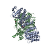
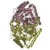
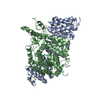
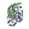
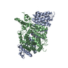
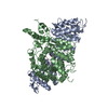
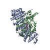
- PDB-6lcu: structure of maltooligosyltrehalose synthase from Arthrobacter ramosus -
+ Open data

- Basic information
| Entry | Database: PDB / ID: 6lcu | ||||||
|---|---|---|---|---|---|---|---|
| Title | structure of maltooligosyltrehalose synthase from Arthrobacter ramosus | ||||||
 Components Components | MTSase | ||||||
 Keywords Keywords | ISOMERASE / maltooligosyltrehalose synthase trehalose | ||||||
| Function / homology |  Function and homology information Function and homology information(1->4)-alpha-D-glucan 1-alpha-D-glucosylmutase / (1,4)-alpha-D-glucan 1-alpha-D-glucosylmutase activity / alpha-glucan catabolic process / trehalose biosynthetic process Similarity search - Function | ||||||
| Biological species |  Arthrobacter ramosus (bacteria) Arthrobacter ramosus (bacteria) | ||||||
| Method |  X-RAY DIFFRACTION / SYNCHROTRON / MOLECULAR REPLACEMENT / Resolution: 2.45 Å X-RAY DIFFRACTION / SYNCHROTRON / MOLECULAR REPLACEMENT / Resolution: 2.45 Å | ||||||
 Authors Authors | Chen, C. / Su, L. / Wu, L. / Zhou, J. / Wu, J. | ||||||
 Citation Citation | Journal: To Be Published Title: structure of maltooligosyltrehalose synthase from Arthrobacter ramosus Authors: Chen, C. / Wu, J. | ||||||
| History |
|
- Structure visualization
| Structure viewer | Molecule: MolmilJmol/JSmol |
|---|
- Downloads & links
-Download
| PDBx/mmCIF format | 6lcu.cif.gz | 369.2 KB | Display | PDBx/mmCIF format |
|---|---|---|---|---|
| PDB format | pdb6lcu.ent.gz | 267.2 KB | Display | PDB format |
| PDBx/mmJSON format | 6lcu.json.gz | Tree view | PDBx/mmJSON format | |
| Others |  Other downloads Other downloads |
-Validation report
| Arichive directory | https://data.pdbj.org/pub/pdb/validation_reports/lc/6lcuftp://data.pdbj.org/pub/pdb/validation_reports/lc/6lcu | HTTPS FTP |
|---|
-Related structure data
| Related structure data |  5zcrS S: Starting model for refinement |
|---|---|
| Similar structure data |


-Links
 PDBj
PDBj

- Assembly
| Deposited unit | 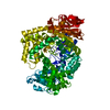
| |||||||||||||||||||||
|---|---|---|---|---|---|---|---|---|---|---|---|---|---|---|---|---|---|---|---|---|---|---|
| 1 |
| |||||||||||||||||||||
| Unit cell |
| |||||||||||||||||||||
| Components on special symmetry positions |
|
-Components
| #1: Protein | Mass: 84905.188 Da / Num. of mol.: 1 Source method: isolated from a genetically manipulated source Source: (gene. exp.) Arthrobacter ramosus (bacteria) / Gene: treYProduction host: References: UniProt: Q9AJN7, (1->4)-alpha-D-glucan 1-alpha-D-glucosylmutase |
|---|---|
| #2: Water | ChemComp-HOH /  Mass: 18.015 Da / Num. of mol.: 575 / Source method: isolated from a natural source / Formula: H2O Mass: 18.015 Da / Num. of mol.: 575 / Source method: isolated from a natural source / Formula: H2O |
-Experimental details
-Experiment
| Experiment | Method: X-RAY DIFFRACTION / Number of used crystals: 1 |
|---|
- Sample preparation
| Crystal | Density Matthews: 6.42 Å3/Da / Density % sol: 80.84 % |
|---|---|
| Crystal grow | Temperature: 289 K / Method: vapor diffusion, hanging drop / Details: Succinic acid |
-Data collection
| Diffraction | Mean temperature: 100 K / Serial crystal experiment: N |
|---|---|
| Diffraction source | Source: SYNCHROTRON / Site: SSRF  / Beamline: BL17U1 / Wavelength: 0.97919 Å / Beamline: BL17U1 / Wavelength: 0.97919 Å |
| Detector | Type: DECTRIS EIGER X 16M / Detector: PIXEL / Date: Aug 12, 2019 |
| Radiation | Protocol: SINGLE WAVELENGTH / Monochromatic (M) / Laue (L): M / Scattering type: x-ray |
| Radiation wavelength | Wavelength: 0.97919 Å / Relative weight: 1 |
| Reflection | Resolution: 2.45→50 Å / Num. obs: 81790 / % possible obs: 100 % / Redundancy: 21 % / Biso Wilson estimate: 39.22 Å2 / CC1/2: 0.997 / Net I/σ(I): 1.85 |
| Reflection shell | Resolution: 2.45→2.49 Å / Num. unique obs: 4011 / CC1/2: 0.718 |
- Processing
| Software |
| ||||||||||||||||||||||||||||||||||||||||||||||||||||||||||||||||||||||||||||||||||||||||||||||||||||||||||||||||||||||||||||||||||||||||||||||||||||||||||||||||||||||||||||||||||||||||||||||||||||||||||||||||||
|---|---|---|---|---|---|---|---|---|---|---|---|---|---|---|---|---|---|---|---|---|---|---|---|---|---|---|---|---|---|---|---|---|---|---|---|---|---|---|---|---|---|---|---|---|---|---|---|---|---|---|---|---|---|---|---|---|---|---|---|---|---|---|---|---|---|---|---|---|---|---|---|---|---|---|---|---|---|---|---|---|---|---|---|---|---|---|---|---|---|---|---|---|---|---|---|---|---|---|---|---|---|---|---|---|---|---|---|---|---|---|---|---|---|---|---|---|---|---|---|---|---|---|---|---|---|---|---|---|---|---|---|---|---|---|---|---|---|---|---|---|---|---|---|---|---|---|---|---|---|---|---|---|---|---|---|---|---|---|---|---|---|---|---|---|---|---|---|---|---|---|---|---|---|---|---|---|---|---|---|---|---|---|---|---|---|---|---|---|---|---|---|---|---|---|---|---|---|---|---|---|---|---|---|---|---|---|---|---|---|---|---|
| Refinement | Method to determine structure: MOLECULAR REPLACEMENT Starting model: 5ZCR Resolution: 2.45→50 Å / SU ML: 0.2378 / Cross valid method: FREE R-VALUE / σ(F): 1.34 / Phase error: 17.5798
| ||||||||||||||||||||||||||||||||||||||||||||||||||||||||||||||||||||||||||||||||||||||||||||||||||||||||||||||||||||||||||||||||||||||||||||||||||||||||||||||||||||||||||||||||||||||||||||||||||||||||||||||||||
| Solvent computation | Shrinkage radii: 0.9 Å / VDW probe radii: 1.11 Å | ||||||||||||||||||||||||||||||||||||||||||||||||||||||||||||||||||||||||||||||||||||||||||||||||||||||||||||||||||||||||||||||||||||||||||||||||||||||||||||||||||||||||||||||||||||||||||||||||||||||||||||||||||
| Displacement parameters | Biso mean: 44.98 Å2 | ||||||||||||||||||||||||||||||||||||||||||||||||||||||||||||||||||||||||||||||||||||||||||||||||||||||||||||||||||||||||||||||||||||||||||||||||||||||||||||||||||||||||||||||||||||||||||||||||||||||||||||||||||
| Refinement step | Cycle: LAST / Resolution: 2.45→50 Å
| ||||||||||||||||||||||||||||||||||||||||||||||||||||||||||||||||||||||||||||||||||||||||||||||||||||||||||||||||||||||||||||||||||||||||||||||||||||||||||||||||||||||||||||||||||||||||||||||||||||||||||||||||||
| Refine LS restraints |
| ||||||||||||||||||||||||||||||||||||||||||||||||||||||||||||||||||||||||||||||||||||||||||||||||||||||||||||||||||||||||||||||||||||||||||||||||||||||||||||||||||||||||||||||||||||||||||||||||||||||||||||||||||
| LS refinement shell |
| ||||||||||||||||||||||||||||||||||||||||||||||||||||||||||||||||||||||||||||||||||||||||||||||||||||||||||||||||||||||||||||||||||||||||||||||||||||||||||||||||||||||||||||||||||||||||||||||||||||||||||||||||||
| Refinement TLS params. | Method: refined / Origin x: -93.2257933485 Å / Origin y: 32.5157817655 Å / Origin z: 10.2670959761 Å
| ||||||||||||||||||||||||||||||||||||||||||||||||||||||||||||||||||||||||||||||||||||||||||||||||||||||||||||||||||||||||||||||||||||||||||||||||||||||||||||||||||||||||||||||||||||||||||||||||||||||||||||||||||
| Refinement TLS group | Selection details: all |
