 Movie
Movie Controller

[English] 日本語
 Yorodumi
- PDB-6lcn: Crystal structure of Serine Acetyltransferase from Planctomyces l... -
+ Open data

- Basic information
| Entry | Database: PDB / ID: 6lcn | |||||||||
|---|---|---|---|---|---|---|---|---|---|---|
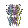
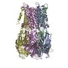
| Title | Crystal structure of Serine Acetyltransferase from Planctomyces limnophilus at 2.15A | |||||||||
 Components Components | Serine O-acetyltransferase | |||||||||
 Keywords Keywords | TRANSFERASE / PSI-Biology / IFN / Structural Genomics | |||||||||
| Function / homology | L-serine O-acetyltransferase activity / Serine acetyltransferase, N-terminal domain superfamily / Serine acetyltransferase, LbH domain / serine O-acetyltransferase / Trimeric LpxA-like superfamily / amino acid biosynthetic process / DI(HYDROXYETHYL)ETHER / Serine O-acetyltransferase Function and homology information Function and homology information | |||||||||
| Biological species |  Planctopirus limnophila DSM 3776 (bacteria) Planctopirus limnophila DSM 3776 (bacteria) | |||||||||
| Method |  X-RAY DIFFRACTION / MOLECULAR REPLACEMENT / Resolution: 2.15 Å X-RAY DIFFRACTION / MOLECULAR REPLACEMENT / Resolution: 2.15 Å | |||||||||
 Authors Authors | Kumar, N. / Singh, R.P. / Singh, A.K. / Kumaran, S. | |||||||||
| Funding support |  India, 2items India, 2items
| |||||||||
 Citation Citation | Journal: To Be Published Title: Understanding Mechanics of competitive-allostery Using Engineered Cysteine Synthase Assembly Authors: Kumar, N. / Singh, R.P. / Kumaran, S. | |||||||||
| History |
|
- Structure visualization
| Structure viewer | Molecule: MolmilJmol/JSmol |
|---|
- Downloads & links
-Download
| PDBx/mmCIF format | 6lcn.cif.gz | 384 KB | Display | PDBx/mmCIF format |
|---|---|---|---|---|
| PDB format | pdb6lcn.ent.gz | 301 KB | Display | PDB format |
| PDBx/mmJSON format | 6lcn.json.gz | Tree view | PDBx/mmJSON format | |
| Others |  Other downloads Other downloads |
-Validation report
| Arichive directory | https://data.pdbj.org/pub/pdb/validation_reports/lc/6lcnftp://data.pdbj.org/pub/pdb/validation_reports/lc/6lcn | HTTPS FTP |
|---|
-Related structure data
| Related structure data |  3f1xS S: Starting model for refinement |
|---|---|
| Similar structure data |


-Links
 PDBj
PDBj


- Assembly
| Deposited unit | 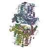
| ||||||||
|---|---|---|---|---|---|---|---|---|---|
| 1 |
| ||||||||
| Unit cell |
|
-Components
| #1: Protein | Mass: 36859.645 Da / Num. of mol.: 6 Source method: isolated from a genetically manipulated source Source: (gene. exp.) Planctopirus limnophila DSM 3776 (bacteria)Strain: DSM 3776 / Gene: Plim_1307 / Production host: #2: Chemical | ChemComp-GOL /   Mass: 92.094 Da / Num. of mol.: 6 / Source method: obtained synthetically / Formula: C3H8O3 / Feature type: SUBJECT OF INVESTIGATION Mass: 92.094 Da / Num. of mol.: 6 / Source method: obtained synthetically / Formula: C3H8O3 / Feature type: SUBJECT OF INVESTIGATION#3: Chemical |   Mass: 35.453 Da / Num. of mol.: 3 / Source method: obtained synthetically / Formula: Cl / Feature type: SUBJECT OF INVESTIGATION Mass: 35.453 Da / Num. of mol.: 3 / Source method: obtained synthetically / Formula: Cl / Feature type: SUBJECT OF INVESTIGATION#4: Chemical | ChemComp-PEG /   Mass: 106.120 Da / Num. of mol.: 4 / Source method: obtained synthetically / Formula: C4H10O3 / Feature type: SUBJECT OF INVESTIGATION Mass: 106.120 Da / Num. of mol.: 4 / Source method: obtained synthetically / Formula: C4H10O3 / Feature type: SUBJECT OF INVESTIGATION#5: Water | ChemComp-HOH / |  Mass: 18.015 Da / Num. of mol.: 766 / Source method: isolated from a natural source / Formula: H2O Mass: 18.015 Da / Num. of mol.: 766 / Source method: isolated from a natural source / Formula: H2OHas ligand of interest | Y | Has protein modification | Y | |
|---|
-Experimental details
-Experiment
| Experiment | Method: X-RAY DIFFRACTION / Number of used crystals: 1 |
|---|
- Sample preparation
| Crystal | Density Matthews: 2.71 Å3/Da / Density % sol: 54.56 % |
|---|---|
| Crystal grow | Temperature: 278 K / Method: vapor diffusion, sitting drop / pH: 6.5 / Details: 0.1M MES pH=6.5, 12% w/v PEG 20,000 |
-Data collection
| Diffraction | Mean temperature: 100 K / Serial crystal experiment: N |
|---|---|
| Diffraction source | Source: ROTATING ANODE / Type: RIGAKU / Wavelength: 1.5417 Å |
| Detector | Type: RIGAKU RAXIS / Detector: IMAGE PLATE / Date: Oct 14, 2017 |
| Radiation | Protocol: SINGLE WAVELENGTH / Monochromatic (M) / Laue (L): M / Scattering type: x-ray |
| Radiation wavelength | Wavelength: 1.5417 Å / Relative weight: 1 |
| Reflection | Resolution: 2.15→47.596 Å / Num. obs: 120745 / % possible obs: 99.64 % / Redundancy: 4.7 % / Biso Wilson estimate: 32.4 Å2 / CC1/2: 0.997 / Rmerge(I) obs: 0.09531 / Rpim(I) all: 0.04956 / Rrim(I) all: 0.1078 / Net I/σ(I): 14.44 |
| Reflection shell | Resolution: 2.15→2.227 Å / Redundancy: 4.5 % / Rmerge(I) obs: 0.742 / Num. unique obs: 11863 / CC1/2: 0.658 / Rpim(I) all: 0.3909 / Rrim(I) all: 0.8414 / % possible all: 99.23 |
- Processing
| Software |
| |||||||||||||||||||||||||||||||||||||||||||||||||||||||||||||||||||||||||||||||||||||||||||||||||||||||||||||||||||||||||||||||||||||||||||||||||||||||||||||||||||||||||||||||||||||||||||||||||||||||||||||||||||||||||||||||||||||||
|---|---|---|---|---|---|---|---|---|---|---|---|---|---|---|---|---|---|---|---|---|---|---|---|---|---|---|---|---|---|---|---|---|---|---|---|---|---|---|---|---|---|---|---|---|---|---|---|---|---|---|---|---|---|---|---|---|---|---|---|---|---|---|---|---|---|---|---|---|---|---|---|---|---|---|---|---|---|---|---|---|---|---|---|---|---|---|---|---|---|---|---|---|---|---|---|---|---|---|---|---|---|---|---|---|---|---|---|---|---|---|---|---|---|---|---|---|---|---|---|---|---|---|---|---|---|---|---|---|---|---|---|---|---|---|---|---|---|---|---|---|---|---|---|---|---|---|---|---|---|---|---|---|---|---|---|---|---|---|---|---|---|---|---|---|---|---|---|---|---|---|---|---|---|---|---|---|---|---|---|---|---|---|---|---|---|---|---|---|---|---|---|---|---|---|---|---|---|---|---|---|---|---|---|---|---|---|---|---|---|---|---|---|---|---|---|---|---|---|---|---|---|---|---|---|---|---|---|---|---|---|---|---|
| Refinement | Method to determine structure: MOLECULAR REPLACEMENT Starting model: 3F1X Resolution: 2.15→47.596 Å / Cor.coef. Fo:Fc: 0.959 / Cor.coef. Fo:Fc free: 0.939 / SU B: 5.78 / SU ML: 0.143 / Cross valid method: FREE R-VALUE / ESU R: 0.204 / ESU R Free: 0.172 Details: Hydrogens have been added in their riding positions
| |||||||||||||||||||||||||||||||||||||||||||||||||||||||||||||||||||||||||||||||||||||||||||||||||||||||||||||||||||||||||||||||||||||||||||||||||||||||||||||||||||||||||||||||||||||||||||||||||||||||||||||||||||||||||||||||||||||||
| Solvent computation | Ion probe radii: 0.8 Å / Shrinkage radii: 0.8 Å / VDW probe radii: 1.2 Å | |||||||||||||||||||||||||||||||||||||||||||||||||||||||||||||||||||||||||||||||||||||||||||||||||||||||||||||||||||||||||||||||||||||||||||||||||||||||||||||||||||||||||||||||||||||||||||||||||||||||||||||||||||||||||||||||||||||||
| Displacement parameters | Biso mean: 38.87 Å2
| |||||||||||||||||||||||||||||||||||||||||||||||||||||||||||||||||||||||||||||||||||||||||||||||||||||||||||||||||||||||||||||||||||||||||||||||||||||||||||||||||||||||||||||||||||||||||||||||||||||||||||||||||||||||||||||||||||||||
| Refinement step | Cycle: LAST / Resolution: 2.15→47.596 Å
| |||||||||||||||||||||||||||||||||||||||||||||||||||||||||||||||||||||||||||||||||||||||||||||||||||||||||||||||||||||||||||||||||||||||||||||||||||||||||||||||||||||||||||||||||||||||||||||||||||||||||||||||||||||||||||||||||||||||
| Refine LS restraints |
| |||||||||||||||||||||||||||||||||||||||||||||||||||||||||||||||||||||||||||||||||||||||||||||||||||||||||||||||||||||||||||||||||||||||||||||||||||||||||||||||||||||||||||||||||||||||||||||||||||||||||||||||||||||||||||||||||||||||
| LS refinement shell | Refine-ID: X-RAY DIFFRACTION / Total num. of bins used: 20
|
