 Movie
Movie Controller

[English] 日本語
 Yorodumi
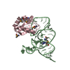
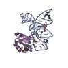
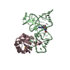
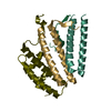
- PDB-6laz: the wildtype SAM-VI riboswitch bound to a N-mustard SAM analog M1 -
+ Open data

- Basic information
| Entry | Database: PDB / ID: 6laz | ||||||
|---|---|---|---|---|---|---|---|
| Title | the wildtype SAM-VI riboswitch bound to a N-mustard SAM analog M1 | ||||||
 Components Components |
| ||||||
 Keywords Keywords | RNA / Riboswitch / SAM / SAM-VI | ||||||
| Function / homology |  Function and homology information Function and homology informationU1 snRNP binding / U1 snRNP / U1 snRNA binding / U4/U6 x U5 tri-snRNP complex / mRNA Splicing - Major Pathway / spliceosomal complex / mRNA splicing, via spliceosome / DNA binding / RNA binding / nucleoplasm ...U1 snRNP binding / U1 snRNP / U1 snRNA binding / U4/U6 x U5 tri-snRNP complex / mRNA Splicing - Major Pathway / spliceosomal complex / mRNA splicing, via spliceosome / DNA binding / RNA binding / nucleoplasm / identical protein binding / nucleus Similarity search - Function | ||||||
| Biological species |  Homo sapiens (human) Homo sapiens (human) Bifidobacterium angulatum (bacteria) Bifidobacterium angulatum (bacteria) | ||||||
| Method |  X-RAY DIFFRACTION / SYNCHROTRON / MOLECULAR REPLACEMENT / Resolution: 2.76 Å X-RAY DIFFRACTION / SYNCHROTRON / MOLECULAR REPLACEMENT / Resolution: 2.76 Å | ||||||
 Authors Authors | Ren, A. / Sun, A. | ||||||
| Funding support |  China, 1items China, 1items
| ||||||
 Citation Citation | Journal: Nat Commun / Year: 2019 Title: SAM-VI riboswitch structure and signature for ligand discrimination. Authors: Sun, A. / Gasser, C. / Li, F. / Chen, H. / Mair, S. / Krasheninina, O. / Micura, R. / Ren, A. | ||||||
| History |
|
- Structure visualization
| Structure viewer | Molecule: MolmilJmol/JSmol |
|---|
- Downloads & links
-Download
| PDBx/mmCIF format | 6laz.cif.gz | 131.8 KB | Display | PDBx/mmCIF format |
|---|---|---|---|---|
| PDB format | pdb6laz.ent.gz | 98 KB | Display | PDB format |
| PDBx/mmJSON format | 6laz.json.gz | Tree view | PDBx/mmJSON format | |
| Others |  Other downloads Other downloads |
-Validation report
| Arichive directory | https://data.pdbj.org/pub/pdb/validation_reports/la/6lazftp://data.pdbj.org/pub/pdb/validation_reports/la/6laz | HTTPS FTP |
|---|
-Related structure data
| Related structure data |  6lasSC  6lauC 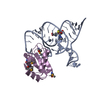 6laxC S: Starting model for refinement C: citing same article ( |
|---|---|
| Similar structure data |


-Links
 PDBj
PDBj


- Assembly
| Deposited unit | 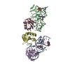
| ||||||||
|---|---|---|---|---|---|---|---|---|---|
| 1 | 
| ||||||||
| 2 | 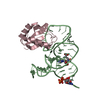
| ||||||||
| 3 | 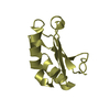
| ||||||||
| Unit cell |
|
-Components
| #1: RNA chain | Mass: 17896.502 Da / Num. of mol.: 2 / Source method: obtained synthetically / Source: (synth.) Bifidobacterium angulatum (bacteria)#2: Protein | Mass: 10796.682 Da / Num. of mol.: 3 / Mutation: Y31H, Q36R, S46K Source method: isolated from a genetically manipulated source Source: (gene. exp.) Homo sapiens (human) / Gene: SNRPA / Production host: #3: Chemical | ChemComp-MG / |   Mass: 24.305 Da / Num. of mol.: 1 / Source method: obtained synthetically / Formula: Mg / Feature type: SUBJECT OF INVESTIGATION Mass: 24.305 Da / Num. of mol.: 1 / Source method: obtained synthetically / Formula: Mg / Feature type: SUBJECT OF INVESTIGATION#4: Chemical | 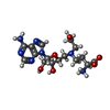  Mass: 411.413 Da / Num. of mol.: 2 / Source method: obtained synthetically / Formula: C16H25N7O6 / Feature type: SUBJECT OF INVESTIGATION Mass: 411.413 Da / Num. of mol.: 2 / Source method: obtained synthetically / Formula: C16H25N7O6 / Feature type: SUBJECT OF INVESTIGATION#5: Water | ChemComp-HOH / |  Mass: 18.015 Da / Num. of mol.: 18 / Source method: isolated from a natural source / Formula: H2O Mass: 18.015 Da / Num. of mol.: 18 / Source method: isolated from a natural source / Formula: H2OHas ligand of interest | Y | Has protein modification | N | |
|---|
-Experimental details
-Experiment
| Experiment | Method: X-RAY DIFFRACTION / Number of used crystals: 1 |
|---|
- Sample preparation
| Crystal | Density Matthews: 3.17 Å3/Da / Density % sol: 61.19 % |
|---|---|
| Crystal grow | Temperature: 289 K / Method: vapor diffusion, sitting drop Details: 0.1 M sodium acetate trihydrate pH 4.6, 10% w/v polyethylene glycol 4,000 |
-Data collection
| Diffraction | Mean temperature: 80 K / Serial crystal experiment: N | ||||||||||||||||||||||||||||||
|---|---|---|---|---|---|---|---|---|---|---|---|---|---|---|---|---|---|---|---|---|---|---|---|---|---|---|---|---|---|---|---|
| Diffraction source | Source: SYNCHROTRON / Site: SSRF / Beamline: BL17U1 / Wavelength: 1.102 Å | ||||||||||||||||||||||||||||||
| Detector | Type: DECTRIS PILATUS3 6M / Detector: PIXEL / Date: Jul 22, 2019 | ||||||||||||||||||||||||||||||
| Radiation | Protocol: SINGLE WAVELENGTH / Monochromatic (M) / Laue (L): M / Scattering type: x-ray | ||||||||||||||||||||||||||||||
| Radiation wavelength | Wavelength: 1.102 Å / Relative weight: 1 | ||||||||||||||||||||||||||||||
| Reflection | Resolution: 2.76→42.67 Å / Num. obs: 20968 / % possible obs: 94.8 % / Redundancy: 6.9 % / Biso Wilson estimate: 73.11 Å2 / CC1/2: 0.998 / Rmerge(I) obs: 0.075 / Rpim(I) all: 0.031 / Rrim(I) all: 0.081 / Net I/σ(I): 15.9 | ||||||||||||||||||||||||||||||
| Reflection shell | Diffraction-ID: 1
|
- Processing
| Software |
| ||||||||||||||||||||||||||||||||||||||||||||||||
|---|---|---|---|---|---|---|---|---|---|---|---|---|---|---|---|---|---|---|---|---|---|---|---|---|---|---|---|---|---|---|---|---|---|---|---|---|---|---|---|---|---|---|---|---|---|---|---|---|---|
| Refinement | Method to determine structure: MOLECULAR REPLACEMENT Starting model: 6LAS Resolution: 2.76→42.67 Å / SU ML: 0.35 / Cross valid method: THROUGHOUT / σ(F): 1.39 / Phase error: 26.38
| ||||||||||||||||||||||||||||||||||||||||||||||||
| Solvent computation | Shrinkage radii: 0.9 Å / VDW probe radii: 1.11 Å | ||||||||||||||||||||||||||||||||||||||||||||||||
| Displacement parameters | Biso max: 167.21 Å2 / Biso mean: 79.1927 Å2 / Biso min: 42.83 Å2 | ||||||||||||||||||||||||||||||||||||||||||||||||
| Refinement step | Cycle: final / Resolution: 2.76→42.67 Å
| ||||||||||||||||||||||||||||||||||||||||||||||||
| Refine LS restraints |
| ||||||||||||||||||||||||||||||||||||||||||||||||
| LS refinement shell | Refine-ID: X-RAY DIFFRACTION / Rfactor Rfree error: 0
|
