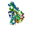
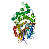
 Movie
Movie Controller


+ Open data

- Basic information
| Entry | Database: PDB / ID: 6l9x | ||||||
|---|---|---|---|---|---|---|---|
| Title | Xenons in frog EPDR1 | ||||||
 Components Components | Ependymin-related 1 | ||||||
 Keywords Keywords | UNKNOWN FUNCTION / EPDR1 / Xenon | ||||||
| Function / homology |  Function and homology information Function and homology informationlysosomal lumen / cell-matrix adhesion / lysosome / calcium ion binding / extracellular region Similarity search - Function | ||||||
| Biological species | |||||||
| Method |  X-RAY DIFFRACTION / SYNCHROTRON / SAD / Resolution: 2.9 Å X-RAY DIFFRACTION / SYNCHROTRON / SAD / Resolution: 2.9 Å | ||||||
 Authors Authors | Park, S. | ||||||
| Funding support |  Korea, Republic Of, 1items Korea, Republic Of, 1items
| ||||||
 Citation Citation | Journal: Crystals / Year: 2020 Title: De novo Phasing Xenons Observed in the Frog Ependymin-Related Protein Authors: Park, S. | ||||||
| History |
|
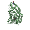
- Structure visualization
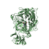
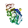
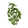
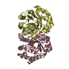
| Structure viewer | Molecule: MolmilJmol/JSmol |
|---|
- Downloads & links
-Download
| PDBx/mmCIF format | 6l9x.cif.gz | 52.9 KB | Display | PDBx/mmCIF format |
|---|---|---|---|---|
| PDB format | pdb6l9x.ent.gz | 36.8 KB | Display | PDB format |
| PDBx/mmJSON format | 6l9x.json.gz | Tree view | PDBx/mmJSON format | |
| Others |  Other downloads Other downloads |
-Validation report
| Arichive directory | https://data.pdbj.org/pub/pdb/validation_reports/l9/6l9xftp://data.pdbj.org/pub/pdb/validation_reports/l9/6l9x | HTTPS FTP |
|---|
-Related structure data
| Similar structure data |
|---|


-Links
 PDBj
PDBj
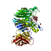
- Assembly
| Deposited unit | 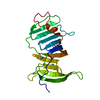
| ||||||||
|---|---|---|---|---|---|---|---|---|---|
| 1 | 
| ||||||||
| Unit cell |
| ||||||||
| Components on special symmetry positions |
|
-Components
| #1: Protein | Mass: 22836.588 Da / Num. of mol.: 1 Source method: isolated from a genetically manipulated source Source: (gene. exp.) Gene: epdr1 / Production host:   Spodoptera frugiperda (fall armyworm) / References: UniProt: F6VRB7 Spodoptera frugiperda (fall armyworm) / References: UniProt: F6VRB7 | ||||
|---|---|---|---|---|---|
| #2: Chemical | ChemComp-XE /   Mass: 131.293 Da / Num. of mol.: 4 / Source method: obtained synthetically / Formula: Xe / Feature type: SUBJECT OF INVESTIGATION Mass: 131.293 Da / Num. of mol.: 4 / Source method: obtained synthetically / Formula: Xe / Feature type: SUBJECT OF INVESTIGATIONHas ligand of interest | Y | Has protein modification | Y | |
-Experimental details
-Experiment
| Experiment | Method: X-RAY DIFFRACTION / Number of used crystals: 1 |
|---|
- Sample preparation
| Crystal | Density Matthews: 2.96 Å3/Da / Density % sol: 58.5 % |
|---|---|
| Crystal grow | Temperature: 298 K / Method: vapor diffusion, hanging drop Details: PEG 8000, 0.2M calcium acetate, 0.1M sodium cacodylate pH 6.5 |
-Data collection
| Diffraction | Mean temperature: 100 K / Serial crystal experiment: N |
|---|---|
| Diffraction source | Source: SYNCHROTRON / Site: PAL/PLS / Beamline: 7A (6B, 6C1) / Wavelength: 1.54 Å |
| Detector | Type: ADSC QUANTUM 270 / Detector: CCD / Date: Jul 25, 2018 |
| Radiation | Protocol: SINGLE WAVELENGTH / Monochromatic (M) / Laue (L): M / Scattering type: x-ray |
| Radiation wavelength | Wavelength: 1.54 Å / Relative weight: 1 |
| Reflection | Resolution: 2.9→50 Å / Num. obs: 6390 / % possible obs: 100 % / Redundancy: 20 % / CC1/2: 0.976 / Rmerge(I) obs: 0.097 / Net I/σ(I): 71 |
| Reflection shell | Resolution: 2.9→2.95 Å / Rmerge(I) obs: 0.582 / Num. unique obs: 2312 / CC1/2: 0.976 |
- Processing
| Software |
| ||||||||||||||||||||||||||||||||||||||||||||||||||||||||||||
|---|---|---|---|---|---|---|---|---|---|---|---|---|---|---|---|---|---|---|---|---|---|---|---|---|---|---|---|---|---|---|---|---|---|---|---|---|---|---|---|---|---|---|---|---|---|---|---|---|---|---|---|---|---|---|---|---|---|---|---|---|---|
| Refinement | Method to determine structure: SAD / Resolution: 2.9→50 Å / Cor.coef. Fo:Fc: 0.93 / Cor.coef. Fo:Fc free: 0.868 / SU B: 16.812 / SU ML: 0.325 / Cross valid method: THROUGHOUT / σ(F): 0 / ESU R Free: 0.454 Details: HYDROGENS HAVE BEEN ADDED IN THE RIDING POSITIONS U VALUES : REFINED INDIVIDUALLY
| ||||||||||||||||||||||||||||||||||||||||||||||||||||||||||||
| Solvent computation | Ion probe radii: 0.8 Å / Shrinkage radii: 0.8 Å / VDW probe radii: 1.2 Å | ||||||||||||||||||||||||||||||||||||||||||||||||||||||||||||
| Displacement parameters | Biso max: 185.66 Å2 / Biso mean: 63.6 Å2 / Biso min: 39.49 Å2
| ||||||||||||||||||||||||||||||||||||||||||||||||||||||||||||
| Refinement step | Cycle: final / Resolution: 2.9→50 Å
| ||||||||||||||||||||||||||||||||||||||||||||||||||||||||||||
| Refine LS restraints |
| ||||||||||||||||||||||||||||||||||||||||||||||||||||||||||||
| LS refinement shell | Resolution: 2.903→2.978 Å / Rfactor Rfree error: 0 / Total num. of bins used: 20
|
