 Movie
Movie Controller

[English] 日本語
 Yorodumi
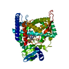
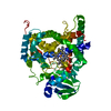
- PDB-6l8j: Crystal structure of CYP97A3 mutant S290D/W300L/S304V in complex ... -
+ Open data

- Basic information
| Entry | Database: PDB / ID: 6l8j | ||||||
|---|---|---|---|---|---|---|---|
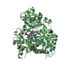
| Title | Crystal structure of CYP97A3 mutant S290D/W300L/S304V in complex with retinal | ||||||
 Components Components | Protein LUTEIN DEFICIENT 5, chloroplastic | ||||||
 Keywords Keywords | OXIDOREDUCTASE / lutein biosynthesis / photosynthesis / monooxygenase / carotenoid / P450 | ||||||
| Function / homology |  Function and homology information Function and homology informationbeta-carotene 3-hydroxylase activity / xanthophyll biosynthetic process / Oxidoreductases; Acting on paired donors, with incorporation or reduction of molecular oxygen / carotenoid biosynthetic process / chloroplast envelope / oxidoreductase activity, acting on paired donors, with incorporation or reduction of molecular oxygen / chloroplast / iron ion binding / heme binding Similarity search - Function | ||||||
| Biological species |  | ||||||
| Method |  X-RAY DIFFRACTION / SYNCHROTRON / MOLECULAR REPLACEMENT / Resolution: 2.399 Å X-RAY DIFFRACTION / SYNCHROTRON / MOLECULAR REPLACEMENT / Resolution: 2.399 Å | ||||||
 Authors Authors | Niu, G. / Guo, Q. / Liu, L. | ||||||
 Citation Citation | Journal: Proc.Natl.Acad.Sci.USA / Year: 2020 Title: Structural basis for plant lutein biosynthesis from alpha-carotene. Authors: Niu, G. / Guo, Q. / Wang, J. / Zhao, S. / He, Y. / Liu, L. | ||||||
| History |
|
- Structure visualization
| Structure viewer | Molecule: MolmilJmol/JSmol |
|---|
- Downloads & links
-Download
| PDBx/mmCIF format | 6l8j.cif.gz | 110 KB | Display | PDBx/mmCIF format |
|---|---|---|---|---|
| PDB format | pdb6l8j.ent.gz | 78.3 KB | Display | PDB format |
| PDBx/mmJSON format | 6l8j.json.gz | Tree view | PDBx/mmJSON format | |
| Others |  Other downloads Other downloads |
-Validation report
| Arichive directory | https://data.pdbj.org/pub/pdb/validation_reports/l8/6l8jftp://data.pdbj.org/pub/pdb/validation_reports/l8/6l8j | HTTPS FTP |
|---|
-Related structure data
| Related structure data |  6j94C  6j95C  6l8hC  6l8iC  4kf2S S: Starting model for refinement C: citing same article ( |
|---|---|
| Similar structure data |


-Links
 PDBj
PDBj


- Assembly
| Deposited unit | 
| ||||||||
|---|---|---|---|---|---|---|---|---|---|
| 1 |
| ||||||||
| Unit cell |
|
-Components
| #1: Protein | Mass: 58430.836 Da / Num. of mol.: 1 / Mutation: S290D,W300L,S304V Source method: isolated from a genetically manipulated source Source: (gene. exp.)  References: UniProt: Q93VK5, Oxidoreductases; Acting on paired donors, with incorporation or reduction of molecular oxygen |
|---|---|
| #2: Chemical | ChemComp-HEM /   Mass: 616.487 Da / Num. of mol.: 1 / Source method: obtained synthetically / Formula: C34H32FeN4O4 / Feature type: SUBJECT OF INVESTIGATION Mass: 616.487 Da / Num. of mol.: 1 / Source method: obtained synthetically / Formula: C34H32FeN4O4 / Feature type: SUBJECT OF INVESTIGATION |
| #3: Chemical | ChemComp-RET /   Mass: 284.436 Da / Num. of mol.: 1 / Source method: obtained synthetically / Formula: C20H28O Mass: 284.436 Da / Num. of mol.: 1 / Source method: obtained synthetically / Formula: C20H28O |
| #4: Water | ChemComp-HOH /  Mass: 18.015 Da / Num. of mol.: 176 / Source method: isolated from a natural source / Formula: H2O Mass: 18.015 Da / Num. of mol.: 176 / Source method: isolated from a natural source / Formula: H2O |
| Has ligand of interest | Y |
-Experimental details
-Experiment
| Experiment | Method: X-RAY DIFFRACTION / Number of used crystals: 1 |
|---|
- Sample preparation
| Crystal | Density Matthews: 2.26 Å3/Da / Density % sol: 45.59 % |
|---|---|
| Crystal grow | Temperature: 289 K / Method: vapor diffusion, sitting drop / Details: potassium sulfate, PEG 3350 |
-Data collection
| Diffraction | Mean temperature: 100 K / Serial crystal experiment: N |
|---|---|
| Diffraction source | Source: SYNCHROTRON / Site: SSRF  / Beamline: BL19U1 / Wavelength: 0.979 Å / Beamline: BL19U1 / Wavelength: 0.979 Å |
| Detector | Type: DECTRIS PILATUS3 6M / Detector: PIXEL / Date: Nov 1, 2016 |
| Radiation | Protocol: SINGLE WAVELENGTH / Monochromatic (M) / Laue (L): M / Scattering type: x-ray |
| Radiation wavelength | Wavelength: 0.979 Å / Relative weight: 1 |
| Reflection | Resolution: 2.399→50 Å / Num. obs: 21298 / % possible obs: 99.4 % / Redundancy: 7.2 % / Biso Wilson estimate: 38.94 Å2 / CC1/2: 0.933 / Rmerge(I) obs: 0.136 / Rpim(I) all: 0.054 / Net I/σ(I): 13 |
| Reflection shell | Resolution: 2.4→2.49 Å / Redundancy: 7.3 % / Num. unique obs: 2077 / CC1/2: 0.793 / Rpim(I) all: 0.347 / % possible all: 99.7 |
- Processing
| Software |
| ||||||||||||||||||||||||||||||||||||||||||||||||||||||
|---|---|---|---|---|---|---|---|---|---|---|---|---|---|---|---|---|---|---|---|---|---|---|---|---|---|---|---|---|---|---|---|---|---|---|---|---|---|---|---|---|---|---|---|---|---|---|---|---|---|---|---|---|---|---|---|
| Refinement | Method to determine structure: MOLECULAR REPLACEMENT Starting model: 4KF2 Resolution: 2.399→43.626 Å / SU ML: 0.28 / Cross valid method: THROUGHOUT / σ(F): 1.33 / Phase error: 25.58
| ||||||||||||||||||||||||||||||||||||||||||||||||||||||
| Solvent computation | Shrinkage radii: 0.9 Å / VDW probe radii: 1.11 Å | ||||||||||||||||||||||||||||||||||||||||||||||||||||||
| Displacement parameters | Biso max: 84.02 Å2 / Biso mean: 43.7637 Å2 / Biso min: 33.24 Å2 | ||||||||||||||||||||||||||||||||||||||||||||||||||||||
| Refinement step | Cycle: final / Resolution: 2.399→43.626 Å
| ||||||||||||||||||||||||||||||||||||||||||||||||||||||
| Refine LS restraints |
| ||||||||||||||||||||||||||||||||||||||||||||||||||||||
| LS refinement shell | Refine-ID: X-RAY DIFFRACTION / Rfactor Rfree error: 0
|
