 Movie
Movie Controller


+ Open data

- Basic information
| Entry | Database: PDB / ID: 6kvi | |||||||||||||||||||||||||||
|---|---|---|---|---|---|---|---|---|---|---|---|---|---|---|---|---|---|---|---|---|---|---|---|---|---|---|---|---|
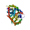
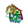
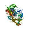
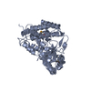
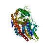
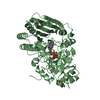
| Title | Crystal structure of UDP-SrUGT76G1 | |||||||||||||||||||||||||||
 Components Components | UDP-glycosyltransferase 76G1 | |||||||||||||||||||||||||||
 Keywords Keywords | TRANSFERASE / glycosyltransferase / diterpenoid / Stevia rebaudiana / natural sweeteners / steviol glycosides (SGs) | |||||||||||||||||||||||||||
| Function / homology |  Function and homology information Function and homology informationsteviolbioside glucosyltransferase activity (rebaudioside B forming) / stevioside glucosyltransferase activity (rebaudioside A forming) / quercetin 3-O-glucosyltransferase activity / quercetin 7-O-glucosyltransferase activity / UDP-glucosyltransferase activity / Transferases; Glycosyltransferases; Hexosyltransferases Similarity search - Function | |||||||||||||||||||||||||||
| Biological species |  Stevia rebaudiana (plant) Stevia rebaudiana (plant) | |||||||||||||||||||||||||||
| Method |  X-RAY DIFFRACTION / SYNCHROTRON / MOLECULAR REPLACEMENT / Resolution: 2.598 Å X-RAY DIFFRACTION / SYNCHROTRON / MOLECULAR REPLACEMENT / Resolution: 2.598 Å | |||||||||||||||||||||||||||
 Authors Authors | Li, J.X. / Liu, Z.F. / Wang, Y. / Zhang, P. | |||||||||||||||||||||||||||
| Funding support |  China, 8items China, 8items
| |||||||||||||||||||||||||||
 Citation Citation | Journal: Plant Commun. / Year: 2020 Title: Structural Insights into the Catalytic Mechanism of a Plant Diterpene Glycosyltransferase SrUGT76G1. Authors: Liu, Z. / Li, J. / Sun, Y. / Zhang, P. / Wang, Y. | |||||||||||||||||||||||||||
| History |
|
- Structure visualization
| Structure viewer | Molecule: MolmilJmol/JSmol |
|---|
- Downloads & links
-Download
| PDBx/mmCIF format | 6kvi.cif.gz | 105.1 KB | Display | PDBx/mmCIF format |
|---|---|---|---|---|
| PDB format | pdb6kvi.ent.gz | 78.3 KB | Display | PDB format |
| PDBx/mmJSON format | 6kvi.json.gz | Tree view | PDBx/mmJSON format | |
| Others |  Other downloads Other downloads |
-Validation report
| Arichive directory | https://data.pdbj.org/pub/pdb/validation_reports/kv/6kviftp://data.pdbj.org/pub/pdb/validation_reports/kv/6kvi | HTTPS FTP |
|---|
-Related structure data
| Related structure data |  6kvjC 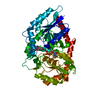 6kvkC 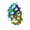 6kvlC 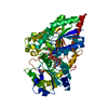 2vceS S: Starting model for refinement C: citing same article ( |
|---|---|
| Similar structure data |


-Links
 PDBj
PDBj


- Assembly
| Deposited unit | 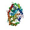
| ||||||||
|---|---|---|---|---|---|---|---|---|---|
| 1 |
| ||||||||
| Unit cell |
|
-Components
| #1: Protein | Mass: 52406.461 Da / Num. of mol.: 1 Source method: isolated from a genetically manipulated source Source: (gene. exp.) Stevia rebaudiana (plant) / Gene: UGT76G1 / Production host:  References: UniProt: Q6VAB4, Transferases; Glycosyltransferases; Hexosyltransferases |
|---|---|
| #2: Chemical | ChemComp-UDP /   Type: RNA linking / Mass: 404.161 Da / Num. of mol.: 1 / Source method: obtained synthetically / Formula: C9H14N2O12P2 / Feature type: SUBJECT OF INVESTIGATION / Comment: UDP*YM Type: RNA linking / Mass: 404.161 Da / Num. of mol.: 1 / Source method: obtained synthetically / Formula: C9H14N2O12P2 / Feature type: SUBJECT OF INVESTIGATION / Comment: UDP*YM |
| #3: Water | ChemComp-HOH /  Mass: 18.015 Da / Num. of mol.: 93 / Source method: isolated from a natural source / Formula: H2O Mass: 18.015 Da / Num. of mol.: 93 / Source method: isolated from a natural source / Formula: H2O |
| Has ligand of interest | Y |
-Experimental details
-Experiment
| Experiment | Method: X-RAY DIFFRACTION / Number of used crystals: 1 |
|---|
- Sample preparation
| Crystal | Density Matthews: 2.59 Å3/Da / Density % sol: 52.57 % |
|---|---|
| Crystal grow | Temperature: 293.15 K / Method: vapor diffusion, sitting drop Details: 18% (vol/vol) 2-propanol, 0.1 M sodium citrate tribasic dihydrate, pH 5.5, 20% (wt/vol) pEG4000 |
-Data collection
| Diffraction | Mean temperature: 100 K / Serial crystal experiment: N |
|---|---|
| Diffraction source | Source: SYNCHROTRON / Site: SSRF / Beamline: BL19U1 / Wavelength: 0.9798 Å |
| Detector | Type: DECTRIS PILATUS 6M / Detector: PIXEL / Date: Sep 28, 2018 |
| Radiation | Protocol: SINGLE WAVELENGTH / Monochromatic (M) / Laue (L): M / Scattering type: x-ray |
| Radiation wavelength | Wavelength: 0.9798 Å / Relative weight: 1 |
| Reflection | Resolution: 2.598→43.228 Å / Num. obs: 15678 / % possible obs: 99.3 % / Redundancy: 6 % / Rrim(I) all: 0.147 / Net I/σ(I): 12.2 |
| Reflection shell | Resolution: 2.598→2.691 Å / Num. unique obs: 1344 / CC1/2: 0.785 |
- Processing
| Software |
| ||||||||||||||||||||||||||||||||||||||||||
|---|---|---|---|---|---|---|---|---|---|---|---|---|---|---|---|---|---|---|---|---|---|---|---|---|---|---|---|---|---|---|---|---|---|---|---|---|---|---|---|---|---|---|---|
| Refinement | Method to determine structure: MOLECULAR REPLACEMENT Starting model: 2vce Resolution: 2.598→43.228 Å / SU ML: 0.37 / Cross valid method: THROUGHOUT / σ(F): 1.34 / Phase error: 24.01 / Stereochemistry target values: ML
| ||||||||||||||||||||||||||||||||||||||||||
| Solvent computation | Shrinkage radii: 0.9 Å / VDW probe radii: 1.11 Å / Solvent model: FLAT BULK SOLVENT MODEL | ||||||||||||||||||||||||||||||||||||||||||
| Displacement parameters | Biso max: 106.16 Å2 / Biso mean: 42.811 Å2 / Biso min: 16.58 Å2 | ||||||||||||||||||||||||||||||||||||||||||
| Refinement step | Cycle: final / Resolution: 2.598→43.228 Å
| ||||||||||||||||||||||||||||||||||||||||||
| Refine LS restraints |
| ||||||||||||||||||||||||||||||||||||||||||
| LS refinement shell | Refine-ID: X-RAY DIFFRACTION / Rfactor Rfree error: 0
|
