Movie
Movie Controller


+ Open data

- Basic information
| Entry | Database: PDB / ID: 6kv5 | |||||||||
|---|---|---|---|---|---|---|---|---|---|---|
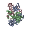
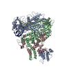
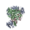
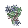
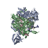
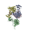
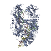
| Title | Structure of influenza D virus apo polymerase | |||||||||
 Components Components |
| |||||||||
 Keywords Keywords | VIRAL PROTEIN / influenza D virus / polymerase / cryo-EM | |||||||||
| Function / homology |  Function and homology information Function and homology informationcap snatching / symbiont-mediated suppression of host mRNA transcription via inhibition of RNA polymerase II activity / host cell mitochondrion / 7-methylguanosine mRNA capping / virion component / symbiont-mediated suppression of host gene expression / RNA-directed RNA polymerase / viral RNA genome replication / nucleotide binding / RNA-directed RNA polymerase activity ...cap snatching / symbiont-mediated suppression of host mRNA transcription via inhibition of RNA polymerase II activity / host cell mitochondrion / 7-methylguanosine mRNA capping / virion component / symbiont-mediated suppression of host gene expression / RNA-directed RNA polymerase / viral RNA genome replication / nucleotide binding / RNA-directed RNA polymerase activity / DNA-templated transcription / RNA binding Similarity search - Function | |||||||||
| Biological species |  Influenza D virus Influenza D virus | |||||||||
| Method | ELECTRON MICROSCOPY / single particle reconstruction / cryo EM / Resolution: 4.6 Å | |||||||||
 Authors Authors | Peng, Q. / Peng, R. / Qi, J. / Gao, G.F. / Shi, Y. | |||||||||
| Funding support |  China, 1items China, 1items
| |||||||||
 Citation Citation | Journal: Nat Microbiol / Year: 2019 Title: Structural insight into RNA synthesis by influenza D polymerase. Authors: Qi Peng / Yuqian Liu / Ruchao Peng / Min Wang / Wei Yang / Hao Song / Yuhai Chen / Sheng Liu / Min Han / Xinzheng Zhang / Peiyi Wang / Jinghua Yan / Buchang Zhang / Jianxun Qi / Tao Deng / ...Authors: Qi Peng / Yuqian Liu / Ruchao Peng / Min Wang / Wei Yang / Hao Song / Yuhai Chen / Sheng Liu / Min Han / Xinzheng Zhang / Peiyi Wang / Jinghua Yan / Buchang Zhang / Jianxun Qi / Tao Deng / George F Gao / Yi Shi / Abstract: The influenza virus polymerase uses capped RNA primers to initiate transcription, and a combination of terminal and internal de novo initiations for the two-step replication process by binding the ...The influenza virus polymerase uses capped RNA primers to initiate transcription, and a combination of terminal and internal de novo initiations for the two-step replication process by binding the conserved viral genomic RNA (vRNA) or complementary RNA (cRNA) promoter. Here, we determined the apo and promoter-bound influenza D polymerase structures using cryo-electron microscopy and found the polymerase has an evolutionarily conserved stable core structure with inherently flexible peripheral domains. Strikingly, two conformations (mode A and B) of the vRNA promoter were observed where the 3'-vRNA end can bind at two different sites, whereas the cRNA promoter only binds in the mode B conformation. Functional studies confirmed the critical role of the mode B conformation for vRNA synthesis via the intermediate cRNA but not for cRNA production, which is mainly regulated by the mode A conformation. Both conformations participate in the regulation of the transcription process. This work advances our understanding of the regulatory mechanisms for the synthesis of different RNA species by influenza virus polymerase and opens new opportunities for antiviral drug design. #1: Journal: Nat Microbiol / Year: 2019Title: Structural insight into RNA synthesis by influenza D polymerase. Authors: Qi Peng / Yuqian Liu / Ruchao Peng / Min Wang / Wei Yang / Hao Song / Yuhai Chen / Sheng Liu / Min Han / Xinzheng Zhang / Peiyi Wang / Jinghua Yan / Buchang Zhang / Jianxun Qi / Tao Deng / ...Authors: Qi Peng / Yuqian Liu / Ruchao Peng / Min Wang / Wei Yang / Hao Song / Yuhai Chen / Sheng Liu / Min Han / Xinzheng Zhang / Peiyi Wang / Jinghua Yan / Buchang Zhang / Jianxun Qi / Tao Deng / George F Gao / Yi Shi / Abstract: The influenza virus polymerase uses capped RNA primers to initiate transcription, and a combination of terminal and internal de novo initiations for the two-step replication process by binding the ...The influenza virus polymerase uses capped RNA primers to initiate transcription, and a combination of terminal and internal de novo initiations for the two-step replication process by binding the conserved viral genomic RNA (vRNA) or complementary RNA (cRNA) promoter. Here, we determined the apo and promoter-bound influenza D polymerase structures using cryo-electron microscopy and found the polymerase has an evolutionarily conserved stable core structure with inherently flexible peripheral domains. Strikingly, two conformations (mode A and B) of the vRNA promoter were observed where the 3'-vRNA end can bind at two different sites, whereas the cRNA promoter only binds in the mode B conformation. Functional studies confirmed the critical role of the mode B conformation for vRNA synthesis via the intermediate cRNA but not for cRNA production, which is mainly regulated by the mode A conformation. Both conformations participate in the regulation of the transcription process. This work advances our understanding of the regulatory mechanisms for the synthesis of different RNA species by influenza virus polymerase and opens new opportunities for antiviral drug design. | |||||||||
| History |
|
- Structure visualization
| Movie |
Movie viewer |
|---|---|
| Structure viewer | Molecule: MolmilJmol/JSmol |
- Downloads & links
-Download
| PDBx/mmCIF format | 6kv5.cif.gz | 319.8 KB | Display | PDBx/mmCIF format |
|---|---|---|---|---|
| PDB format | pdb6kv5.ent.gz | 249.7 KB | Display | PDB format |
| PDBx/mmJSON format | 6kv5.json.gz | Tree view | PDBx/mmJSON format | |
| Others |  Other downloads Other downloads |
-Validation report
| Arichive directory | https://data.pdbj.org/pub/pdb/validation_reports/kv/6kv5ftp://data.pdbj.org/pub/pdb/validation_reports/kv/6kv5 | HTTPS FTP |
|---|
-Related structure data
| Related structure data | 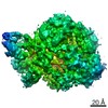 9577MC 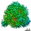 9578C  9579C 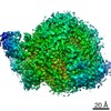 9580C 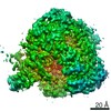 9581C 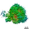 9582C 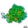 9887C 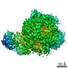 9888C 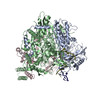 6kujC 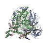 6kukC 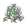 6kupC 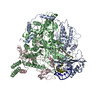 6kurC 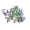 6kutC  6kuuC 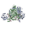 6kuvC M: map data used to model this data C: citing same article ( |
|---|---|
| Similar structure data |


-Links
 PDBj
PDBj- Assembly
| Deposited unit | 
|
|---|---|
| 1 |
|
-Components
| #1: Protein | Mass: 83036.086 Da / Num. of mol.: 1 Source method: isolated from a genetically manipulated source Source: (gene. exp.) Influenza D virus (D/swine/Oklahoma/1334/2011)Strain: D/swine/Oklahoma/1334/2011 / Gene: P3 Production host:  Spodoptera aff. frugiperda 1 BOLD-2017 (butterflies/moths) Spodoptera aff. frugiperda 1 BOLD-2017 (butterflies/moths)References: UniProt: K9LHJ4 |
|---|---|
| #2: Protein | Mass: 86138.844 Da / Num. of mol.: 1 Source method: isolated from a genetically manipulated source Source: (gene. exp.) Influenza D virus (D/swine/Oklahoma/1334/2011)Strain: D/swine/Oklahoma/1334/2011 / Gene: PB1 Production host: Spodoptera aff. frugiperda 1 BOLD-2017 (butterflies/moths)References: UniProt: K9LH03, RNA-directed RNA polymerase |
| #3: Protein | Mass: 88480.484 Da / Num. of mol.: 1 Source method: isolated from a genetically manipulated source Source: (gene. exp.) Influenza D virus (D/swine/Oklahoma/1334/2011)Strain: D/swine/Oklahoma/1334/2011 / Gene: PB2 Production host: Spodoptera aff. frugiperda 1 BOLD-2017 (butterflies/moths)References: UniProt: K9LHF3 |
-Experimental details
-Experiment
| Experiment | Method: ELECTRON MICROSCOPY |
|---|---|
| EM experiment | Aggregation state: PARTICLE / 3D reconstruction method: single particle reconstruction |
- Sample preparation
| Component | Name: Influenza D virus polymerase complex / Type: COMPLEX / Entity ID: all / Source: RECOMBINANT |
|---|---|
| Molecular weight | Value: 0.25 MDa / Experimental value: YES |
| Source (natural) | Organism: Influenza D virus |
| Source (recombinant) | Organism: Spodoptera aff. frugiperda 1 BOLD-2017 (butterflies/moths) |
| Buffer solution | pH: 7.5 |
| Specimen | Embedding applied: NO / Shadowing applied: NO / Staining applied: NO / Vitrification applied: YES |
| Specimen support | Grid material: COPPER / Grid type: Quantifoil R1.2/1.3 |
| Vitrification | Instrument: FEI VITROBOT MARK IV / Cryogen name: ETHANE |
- Electron microscopy imaging
| Experimental equipment |  Model: Titan Krios / Image courtesy: FEI Company |
|---|---|
| Microscopy | Model: FEI TITAN KRIOS |
| Electron gun | Electron source:  FIELD EMISSION GUN / Accelerating voltage: 300 kV / Illumination mode: FLOOD BEAM FIELD EMISSION GUN / Accelerating voltage: 300 kV / Illumination mode: FLOOD BEAM |
| Electron lens | Mode: BRIGHT FIELD / Alignment procedure: COMA FREE |
| Specimen holder | Cryogen: NITROGEN / Specimen holder model: FEI TITAN KRIOS AUTOGRID HOLDER |
| Image recording | Electron dose: 60 e/Å2 / Detector mode: SUPER-RESOLUTION / Film or detector model: GATAN K2 SUMMIT (4k x 4k) |
| EM imaging optics | Energyfilter name: GIF Bioquantum / Energyfilter slit width: 20 eV |
- Processing
| Software | Name: PHENIX / Version: 1.11.1_2575: / Classification: refinement | ||||||||||||||||||||||||||||||||||||
|---|---|---|---|---|---|---|---|---|---|---|---|---|---|---|---|---|---|---|---|---|---|---|---|---|---|---|---|---|---|---|---|---|---|---|---|---|---|
| EM software |
| ||||||||||||||||||||||||||||||||||||
| CTF correction | Type: PHASE FLIPPING AND AMPLITUDE CORRECTION | ||||||||||||||||||||||||||||||||||||
| Symmetry | Point symmetry: C1 (asymmetric) | ||||||||||||||||||||||||||||||||||||
| 3D reconstruction | Resolution: 4.6 Å / Resolution method: FSC 0.143 CUT-OFF / Num. of particles: 108120 / Algorithm: FOURIER SPACE / Symmetry type: POINT | ||||||||||||||||||||||||||||||||||||
| Atomic model building | Protocol: FLEXIBLE FIT / Space: REAL | ||||||||||||||||||||||||||||||||||||
| Atomic model building | PDB-ID: 5D98 Accession code: 5D98 / Source name: PDB / Type: experimental model | ||||||||||||||||||||||||||||||||||||
| Refine LS restraints |
|