| Entry | Database: PDB / ID: 6kth
|
|---|
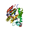
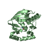
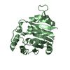
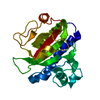
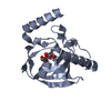
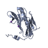
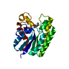
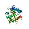
| Title | Crystal structure of Juvenile hormone diol kinase JHDK-L2 from silkworm, Bombyx mori |
|---|
 Components Components | Juvenile hormone diol kinase |
|---|
 Keywords Keywords | HORMONE / kinase / catabolism / EF-hand motif |
|---|
| Function / homology |  Function and homology information Function and homology information
EF hand / EF-hand / Recoverin; domain 1 / EF-hand, calcium binding motif / EF-Hand 1, calcium-binding site / EF-hand calcium-binding domain. / EF-hand calcium-binding domain profile. / EF-hand domain / EF-hand domain pair / Orthogonal Bundle / Mainly AlphaSimilarity search - Domain/homology |
|---|
| Biological species |   Bombyx mori (domestic silkworm) Bombyx mori (domestic silkworm) |
|---|
| Method |  X-RAY DIFFRACTION / SYNCHROTRON / MOLECULAR REPLACEMENT / Resolution: 1.22 Å X-RAY DIFFRACTION / SYNCHROTRON / MOLECULAR REPLACEMENT / Resolution: 1.22 Å |
|---|
 Authors Authors | Zhang, Y.S. / Xu, H.Y. / Wang, Z. / Zhang, L. / Zhao, P. / Guo, P.C. |
|---|
| Funding support |  China, 2items China, 2items | Organization | Grant number | Country |
|---|
| National Natural Science Foundation of China | 31502019 | China | | National Natural Science Foundation of China | 31970468 | China |
|
|---|
 Citation Citation | #1: Journal: J. Mol. Biol. / Year: 2006Title: Structure of the neuronal protein calexcitin suggests a mode of interaction in signalling pathways of learning and memory. Authors: Erskine, P.T. / Beaven, G.D. / Hagan, R. / Findlow, I.S. / Werner, J.M. / Wood, S.P. / Vernon, J. / Giese, K.P. / Fox, G. / Cooper, J.B. |
|---|
| History | | Deposition | Aug 28, 2019 | Deposition site: PDBJ / Processing site: PDBJ |
|---|
| Revision 1.0 | Sep 2, 2020 | Provider: repository / Type: Initial release |
|---|
| Revision 1.1 | Apr 7, 2021 | Group: Database references / Category: citation / citation_author
Item: _citation.country / _citation.journal_abbrev ..._citation.country / _citation.journal_abbrev / _citation.journal_id_ASTM / _citation.journal_id_CSD / _citation.journal_id_ISSN / _citation.journal_volume / _citation.page_first / _citation.page_last / _citation.pdbx_database_id_DOI / _citation.pdbx_database_id_PubMed / _citation.title / _citation.year / _citation_author.name |
|---|
| Revision 1.2 | Nov 22, 2023 | Group: Data collection / Database references ...Data collection / Database references / Derived calculations / Refinement description
Category: atom_type / chem_comp_atom ...atom_type / chem_comp_atom / chem_comp_bond / database_2 / pdbx_initial_refinement_model
Item: _atom_type.pdbx_N_electrons / _atom_type.pdbx_scat_Z ..._atom_type.pdbx_N_electrons / _atom_type.pdbx_scat_Z / _database_2.pdbx_DOI / _database_2.pdbx_database_accession |
|---|
|
|---|
 Movie
Movie Controller

 Yorodumi
 Open data
 Basic information
 Structure visualization
 Downloads & links
 Other downloads
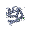

 PDBj
PDBj Assembly
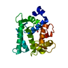


 Mass: 40.078 Da / Num. of mol.: 1 / Source method: obtained synthetically / Formula: Ca
 Mass: 92.094 Da / Num. of mol.: 1 / Source method: obtained synthetically / Formula: C3H8O3
 Mass: 18.015 Da / Num. of mol.: 178 / Source method: isolated from a natural source / Formula: H2O
 Sample preparation
 Processing