 Movie
Movie Controller

[English] 日本語
 Yorodumi
- PDB-6kpd: The crystal structure of the BALDIBIS/IDD9 bound to the homodimer... -
+ Open data

- Basic information
| Entry | Database: PDB / ID: 6kpd | |||||||||
|---|---|---|---|---|---|---|---|---|---|---|
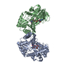
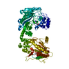
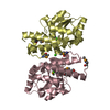
| Title | The crystal structure of the BALDIBIS/IDD9 bound to the homodimeric SCL3 | |||||||||
 Components Components |
| |||||||||
 Keywords Keywords | TRANSCRIPTION / Transcription factor / asymmetric cell division / GRAS / IDD | |||||||||
| Function / homology |  Function and homology information Function and homology informationregulation of meristem growth / response to gibberellin / protein localization to nucleus / sequence-specific DNA binding / DNA-binding transcription factor activity / regulation of DNA-templated transcription / positive regulation of DNA-templated transcription / metal ion binding / nucleus Similarity search - Function | |||||||||
| Biological species |  | |||||||||
| Method |  X-RAY DIFFRACTION / SYNCHROTRON / MOLECULAR REPLACEMENT / Resolution: 3.2 Å X-RAY DIFFRACTION / SYNCHROTRON / MOLECULAR REPLACEMENT / Resolution: 3.2 Å | |||||||||
 Authors Authors | Hirano, Y. / Shimizu, R. / Hakoshima, T. | |||||||||
| Funding support |  Japan, 2items Japan, 2items
| |||||||||
 Citation Citation | Journal: To Be Published Title: Structure of the SCL3 homodimer bound to the BIRD/IDD transcription factor Authors: Hirano, Y. / Shimizu, R. / Nishimura, T. / Morita, M.T. / Hakoshima, T. | |||||||||
| History |
|
- Structure visualization
| Structure viewer | Molecule: MolmilJmol/JSmol |
|---|
- Downloads & links
-Download
| PDBx/mmCIF format | 6kpd.cif.gz | 91.8 KB | Display | PDBx/mmCIF format |
|---|---|---|---|---|
| PDB format | pdb6kpd.ent.gz | 66.7 KB | Display | PDB format |
| PDBx/mmJSON format | 6kpd.json.gz | Tree view | PDBx/mmJSON format | |
| Others |  Other downloads Other downloads |
-Validation report
| Arichive directory | https://data.pdbj.org/pub/pdb/validation_reports/kp/6kpdftp://data.pdbj.org/pub/pdb/validation_reports/kp/6kpd | HTTPS FTP |
|---|
-Related structure data
| Related structure data |  6kpbC  5b3gS C: citing same article ( S: Starting model for refinement |
|---|---|
| Similar structure data |


-Links
 PDBj
PDBj


- Assembly
| Deposited unit | 
| ||||||||||||
|---|---|---|---|---|---|---|---|---|---|---|---|---|---|
| 1 | 
| ||||||||||||
| Unit cell |
|
-Components
| #1: Protein | Mass: 50824.766 Da / Num. of mol.: 1 Source method: isolated from a genetically manipulated source Details: The sample sequence has the N-terminal additional residues (G-P, tag) and a deletion region corresponding to (270-301). These are genetically modified for the recombinant protein expression. Source: (gene. exp.)  |
|---|---|
| #2: Protein/peptide | Mass: 3852.378 Da / Num. of mol.: 1 Source method: isolated from a genetically manipulated source Source: (gene. exp.) |
-Experimental details
-Experiment
| Experiment | Method: X-RAY DIFFRACTION / Number of used crystals: 1 |
|---|
- Sample preparation
| Crystal | Density Matthews: 1.94 Å3/Da / Density % sol: 36.54 % |
|---|---|
| Crystal grow | Temperature: 293 K / Method: vapor diffusion / pH: 6.5 Details: PEG 3350, sodium sulfate, Bis-Tris propane-NaOH (pH 6.5) |
-Data collection
| Diffraction | Mean temperature: 100 K / Serial crystal experiment: N |
|---|---|
| Diffraction source | Source: SYNCHROTRON / Site: SPring-8 / Beamline: BL41XU / Wavelength: 1 Å |
| Detector | Type: DECTRIS PILATUS3 S 6M / Detector: PIXEL / Date: Dec 7, 2017 |
| Radiation | Monochromator: Rotated-inclined double-crystal monochromator , Si (111) Protocol: SINGLE WAVELENGTH / Monochromatic (M) / Laue (L): M / Scattering type: x-ray |
| Radiation wavelength | Wavelength: 1 Å / Relative weight: 1 |
| Reflection | Resolution: 3.2→44.77 Å / Num. obs: 7177 / % possible obs: 99.3 % / Redundancy: 4.2 % / CC1/2: 0.995 / Rmerge(I) obs: 0.115 / Rpim(I) all: 0.061 / Net I/σ(I): 6.7 |
| Reflection shell | Resolution: 3.2→3.42 Å / Redundancy: 2.9 % / Rmerge(I) obs: 0.521 / Mean I/σ(I) obs: 2.9 / Num. unique obs: 1279 / CC1/2: 0.701 / Rpim(I) all: 0.36 / % possible all: 99.1 |
- Processing
| Software |
| ||||||||||||||||||||||||||||
|---|---|---|---|---|---|---|---|---|---|---|---|---|---|---|---|---|---|---|---|---|---|---|---|---|---|---|---|---|---|
| Refinement | Method to determine structure: MOLECULAR REPLACEMENT Starting model: 5B3G Resolution: 3.2→44.77 Å / Cross valid method: FREE R-VALUE
| ||||||||||||||||||||||||||||
| Solvent computation | Shrinkage radii: 0.9 Å / VDW probe radii: 1.11 Å | ||||||||||||||||||||||||||||
| Refinement step | Cycle: LAST / Resolution: 3.2→44.77 Å
| ||||||||||||||||||||||||||||
| LS refinement shell |
|
