 Movie
Movie Controller


+ Open data

- Basic information
| Entry | Database: PDB / ID: 6knf | |||||||||||||||||||||||||||
|---|---|---|---|---|---|---|---|---|---|---|---|---|---|---|---|---|---|---|---|---|---|---|---|---|---|---|---|---|
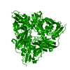
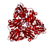
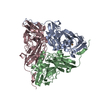
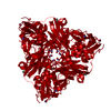
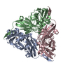
| Title | CryoEM map and model of Nitrite Reductase at pH 6.2 | |||||||||||||||||||||||||||
 Components Components | Copper-containing nitrite reductase | |||||||||||||||||||||||||||
 Keywords Keywords | OXIDOREDUCTASE / Cu containing nitrite reductase | |||||||||||||||||||||||||||
| Function / homology |  Function and homology information Function and homology informationdenitrification pathway / nitrite reductase (NO-forming) / nitrite reductase (NO-forming) activity / nitrate assimilation / periplasmic space / copper ion binding Similarity search - Function | |||||||||||||||||||||||||||
| Biological species |  Achromobacter cycloclastes (bacteria) Achromobacter cycloclastes (bacteria) | |||||||||||||||||||||||||||
| Method | ELECTRON MICROSCOPY / single particle reconstruction / cryo EM / Resolution: 2.99 Å | |||||||||||||||||||||||||||
 Authors Authors | Adachi, N. / Yamaguchi, T. / Moriya, T. / Kawasaki, M. / Koiwai, K. / Shinoda, A. / Yamada, Y. / Yumoto, F. / Kohzuma, T. / Senda, T. | |||||||||||||||||||||||||||
| Funding support |  Japan, 1items Japan, 1items
| |||||||||||||||||||||||||||
 Citation Citation | Journal: J Struct Biol / Year: 2021 Title: 2.85 and 2.99 Å resolution structures of 110 kDa nitrite reductase determined by 200 kV cryogenic electron microscopy. Authors: Naruhiko Adachi / Takahide Yamaguchi / Toshio Moriya / Masato Kawasaki / Kotaro Koiwai / Akira Shinoda / Yusuke Yamada / Fumiaki Yumoto / Takamitsu Kohzuma / Toshiya Senda / Abstract: Cu-containing nitrite reductases (NiRs) are 110 kDa enzymes that play central roles in denitrification. Although the NiRs have been well studied, with over 100 Protein Data Bank entries, such issues ...Cu-containing nitrite reductases (NiRs) are 110 kDa enzymes that play central roles in denitrification. Although the NiRs have been well studied, with over 100 Protein Data Bank entries, such issues as crystal packing, photoreduction, and lack of high pH cases have impeded structural analysis of their catalytic mechanisms. Here we show the cryogenic electron microscopy (cryo-EM) structures of Achromobacter cycloclastes NiR (AcNiR) at pH 6.2 and 8.1. The optimization of 3D-reconstruction parameters achieved 2.99 and 2.85 Å resolution. Comprehensive comparisons with cryo-EM and 56 AcNiR crystal structures suggested crystallographic artifacts in residues 185-215 and His255' due to packing and photoreduction, respectively. We used a newly developed map comparison method to detect structural change around the type 2 Cu site. While the theoretical estimation of coordinate errors of cryo-EM structures remains difficult, combined analysis using X-ray and cryo-EM structures will allow deeper insight into the local structural changes of proteins. | |||||||||||||||||||||||||||
| History |
|
- Structure visualization
| Movie |
Movie viewer |
|---|---|
| Structure viewer | Molecule: MolmilJmol/JSmol |
- Downloads & links
-Download
| PDBx/mmCIF format | 6knf.cif.gz | 177.5 KB | Display | PDBx/mmCIF format |
|---|---|---|---|---|
| PDB format | pdb6knf.ent.gz | 141 KB | Display | PDB format |
| PDBx/mmJSON format | 6knf.json.gz | Tree view | PDBx/mmJSON format | |
| Others |  Other downloads Other downloads |
-Validation report
| Arichive directory | https://data.pdbj.org/pub/pdb/validation_reports/kn/6knfftp://data.pdbj.org/pub/pdb/validation_reports/kn/6knf | HTTPS FTP |
|---|
-Related structure data
| Related structure data | 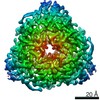 0730MC 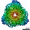 0731C  6kngC M: map data used to model this data C: citing same article ( |
|---|---|
| Similar structure data | |
| EM raw data | EMPIAR-10580 (Title: CryoEM map and model of Nitrite Reductase at pH 6.2 / Data size: 1.2 TB Data #1: CryoEM map and model of Nitrite Reductase at pH 6.2 [micrographs - multiframe]) |


-Links
 PDBj
PDBj

- Assembly
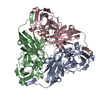
| Deposited unit | 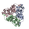
|
|---|---|
| 1 |
|
-Components
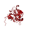
| #1: Protein | Mass: 36621.332 Da / Num. of mol.: 3 / Source method: isolated from a natural source / Source: (natural) Achromobacter cycloclastes (bacteria) / References: UniProt: P25006, nitrite reductase (NO-forming)#2: Chemical | ChemComp-CU /   Mass: 63.546 Da / Num. of mol.: 6 / Source method: obtained synthetically / Formula: Cu / Feature type: SUBJECT OF INVESTIGATION Mass: 63.546 Da / Num. of mol.: 6 / Source method: obtained synthetically / Formula: Cu / Feature type: SUBJECT OF INVESTIGATIONHas ligand of interest | Y | Has protein modification | N | |
|---|
-Experimental details
-Experiment
| Experiment | Method: ELECTRON MICROSCOPY |
|---|---|
| EM experiment | Aggregation state: PARTICLE / 3D reconstruction method: single particle reconstruction |
- Sample preparation
| Component | Name: Nitrite reductase / Type: ORGANELLE OR CELLULAR COMPONENT / Entity ID: #1 / Source: NATURAL |
|---|---|
| Molecular weight | Value: 0.11 MDa / Experimental value: NO |
| Source (natural) | Organism: Achromobacter cycloclastes (bacteria) |
| Buffer solution | pH: 6.2 |
| Buffer component | Conc.: 100 mM / Name: potassium phosphate |
| Specimen | Conc.: 1.1 mg/ml / Embedding applied: NO / Shadowing applied: NO / Staining applied: NO / Vitrification applied: YES / Details: This sample was mono-disperse. |
| Specimen support | Details: The grid was washed by acetone prior to use. / Grid material: COPPER / Grid mesh size: 300 divisions/in. / Grid type: Quantifoil R1.2/1.3 |
| Vitrification | Instrument: FEI VITROBOT MARK IV / Cryogen name: ETHANE / Humidity: 100 % / Chamber temperature: 291 K / Details: Blotting time was 15 seconds |
- Electron microscopy imaging
| Experimental equipment |  Model: Talos Arctica / Image courtesy: FEI Company |
|---|---|
| Microscopy | Model: FEI TALOS ARCTICA |
| Electron gun | Electron source:  FIELD EMISSION GUN / Accelerating voltage: 200 kV / Illumination mode: FLOOD BEAM FIELD EMISSION GUN / Accelerating voltage: 200 kV / Illumination mode: FLOOD BEAM |
| Electron lens | Mode: BRIGHT FIELD / Nominal magnification: 120000 X / Nominal defocus max: 3000 nm / Nominal defocus min: 1000 nm / Cs: 2.7 mm / C2 aperture diameter: 50 µm |
| Specimen holder | Cryogen: NITROGEN |
| Image recording | Electron dose: 50 e/Å2 / Detector mode: COUNTING / Film or detector model: FEI FALCON III (4k x 4k) / Num. of grids imaged: 1 / Num. of real images: 794 |
- Processing
| Software | Name: PHENIX / Version: 1.17.1_3660: / Classification: refinement | ||||||||||||||||||||||||||||||||
|---|---|---|---|---|---|---|---|---|---|---|---|---|---|---|---|---|---|---|---|---|---|---|---|---|---|---|---|---|---|---|---|---|---|
| EM software |
| ||||||||||||||||||||||||||||||||
| CTF correction | Type: PHASE FLIPPING AND AMPLITUDE CORRECTION | ||||||||||||||||||||||||||||||||
| Symmetry | Point symmetry: C3 (3 fold cyclic) | ||||||||||||||||||||||||||||||||
| 3D reconstruction | Resolution: 2.99 Å / Resolution method: FSC 0.143 CUT-OFF / Num. of particles: 121353 / Symmetry type: POINT | ||||||||||||||||||||||||||||||||
| Refine LS restraints |
|
