+
データを開く
-
基本情報
登録情報 | データベース: PDB / ID: 6kj2 | |||||||||||||||
---|---|---|---|---|---|---|---|---|---|---|---|---|---|---|---|---|
タイトル | 200kV MicroED structure of FUS (37-42) SYSGYS solved from single crystal at 0.67 A | |||||||||||||||
![]() | RNA-binding protein FUS | |||||||||||||||
![]() | RNA BINDING PROTEIN / FUS / MicroED / Ultrahigh resolution | |||||||||||||||
機能・相同性 | ![]() membraneless organelle assembly / mRNA stabilization / intracellular membraneless organelle / regulation of RNA splicing / Processing of Capped Intron-Containing Pre-mRNA / postsynaptic cytosol / positive regulation of double-strand break repair via homologous recombination / presynaptic cytosol / mRNA Splicing - Major Pathway / RNA splicing ...membraneless organelle assembly / mRNA stabilization / intracellular membraneless organelle / regulation of RNA splicing / Processing of Capped Intron-Containing Pre-mRNA / postsynaptic cytosol / positive regulation of double-strand break repair via homologous recombination / presynaptic cytosol / mRNA Splicing - Major Pathway / RNA splicing / mRNA 3'-UTR binding / transcription coregulator activity / molecular condensate scaffold activity / GABA-ergic synapse / protein homooligomerization / amyloid fibril formation / transcription coactivator activity / chromatin binding / regulation of transcription by RNA polymerase II / regulation of DNA-templated transcription / glutamatergic synapse / DNA binding / RNA binding / zinc ion binding / nucleoplasm / identical protein binding / nucleus 類似検索 - 分子機能 | |||||||||||||||
生物種 | ![]() | |||||||||||||||
手法 | 電子線結晶学 / クライオ電子顕微鏡法 / 解像度: 0.67 Å | |||||||||||||||
![]() | Zhou, H. / Luo, F. / Luo, Z. / Li, D. / Liu, C. / Li, X. | |||||||||||||||
資金援助 | ![]()
| |||||||||||||||
![]() | ![]() タイトル: Programming Conventional Electron Microscopes for Solving Ultrahigh-Resolution Structures of Small and Macro-Molecules. 著者: Heng Zhou / Feng Luo / Zhipu Luo / Dan Li / Cong Liu / Xueming Li / ![]() 要旨: Microcrystal electron diffraction (MicroED) is becoming a powerful tool in determining the crystal structures of biological macromolecules and small organic compounds. However, wide applications of ...Microcrystal electron diffraction (MicroED) is becoming a powerful tool in determining the crystal structures of biological macromolecules and small organic compounds. However, wide applications of this technique are still limited by the special requirement for radiation-tolerated movie-mode camera and the lack of automated data collection methods. Herein, we develop a stage-camera synchronization scheme to minimize the hardware requirements and enable the use of the conventional electron cryo-microscope with a single-frame CCD camera, which ensures not only the acquisition of ultrahigh-resolution diffraction data but also low cost in practice. This method renders the structure determination of both peptide and small organic compounds at ultrahigh resolution up to ∼0.60 Å with unambiguous assignment of nearly all hydrogen atoms. The present work provides a widely applicable solution for routine structure determination of MicroED and demonstrates the capability of the low-end 120 kV microscope with a CCD camera in solving ultrahigh resolution structures of both organic compounds and biological macromolecules. | |||||||||||||||
履歴 |
|
-
構造の表示
ムービー |
![]() |
---|---|
構造ビューア | 分子: ![]() ![]() |
-
ダウンロードとリンク
-
ダウンロード
PDBx/mmCIF形式 | ![]() | 11.6 KB | 表示 | ![]() |
---|---|---|---|---|
PDB形式 | ![]() | 6.5 KB | 表示 | ![]() |
PDBx/mmJSON形式 | ![]() | ツリー表示 | ![]() | |
その他 | ![]() |
-検証レポート
文書・要旨 | ![]() | 689.1 KB | 表示 | ![]() |
---|---|---|---|---|
文書・詳細版 | ![]() | 688.7 KB | 表示 | |
XML形式データ | ![]() | 6.1 KB | 表示 | |
CIF形式データ | ![]() | 7.9 KB | 表示 | |
アーカイブディレクトリ | ![]() ![]() | HTTPS FTP |
-関連構造データ
関連構造データ | ![]() 0697MC ![]() 0696C ![]() 0698C ![]() 0699C ![]() 6kj1C ![]() 6kj3C ![]() 6kj4C M: このデータのモデリングに利用したマップデータ C: 同じ文献を引用 ( |
---|---|
類似構造データ | 類似検索 - 機能・相同性 ![]() |
-
リンク
-
集合体
登録構造単位 | ![]()
| ||||||||
---|---|---|---|---|---|---|---|---|---|
1 |
| ||||||||
単位格子 |
|
-
要素
#1: タンパク質・ペプチド | 分子量: 662.648 Da / 分子数: 1 / 由来タイプ: 合成 / 由来: (合成) ![]() |
---|---|
#2: 水 | ChemComp-HOH / |
-実験情報
-実験
実験 | 手法: 電子線結晶学 |
---|---|
EM実験 | 試料の集合状態: 3D ARRAY / 3次元再構成法: 電子線結晶学 |
-
試料調製
構成要素 | 名称: FUS LC RAC1 / タイプ: COMPLEX / Entity ID: #1 / 由来: MULTIPLE SOURCES |
---|---|
分子量 | 実験値: NO |
緩衝液 | pH: 7 |
試料 | 包埋: NO / シャドウイング: NO / 染色: NO / 凍結: YES |
急速凍結 | 凍結剤: ETHANE |
-データ収集
実験機器 | ![]() モデル: Tecnai F20 / 画像提供: FEI Company |
---|---|
顕微鏡 | モデル: FEI TECNAI F20 / 日付: 2018年3月16日 |
電子銃 | 電子線源: ![]() |
電子レンズ | モード: DIFFRACTION |
撮影 | 電子線照射量: 0.05 e/Å2 フィルム・検出器のモデル: GATAN ULTRASCAN 4000 (4k x 4k) |
EM回折 | カメラ長: 520 mm |
EM回折 シェル | 解像度: 0.67→0.69 Å / フーリエ空間範囲: 35.97 % / 多重度: 1.99 / 構造因子数: 227 / 位相残差: 1 ° |
EM回折 統計 | フーリエ空間範囲: 58.77 % / 再高解像度: 0.67 Å / 測定した強度の数: 9323 / 構造因子数: 3780 / 位相誤差: 40.93 ° / 位相残差: 40.93 ° / 位相誤差の除外基準: 1 / Rmerge: 0.123 / Rsym: 0.123 |
-
解析
ソフトウェア |
| ||||||||||||||||||||||||
---|---|---|---|---|---|---|---|---|---|---|---|---|---|---|---|---|---|---|---|---|---|---|---|---|---|
EM 3D crystal entity | ∠α: 90 ° / ∠β: 90.06 ° / ∠γ: 90 ° / A: 18.11 Å / B: 4.95 Å / C: 18.66 Å / 空間群名: P1211 / 空間群番号: 4 | ||||||||||||||||||||||||
CTF補正 | タイプ: NONE | ||||||||||||||||||||||||
3次元再構成 | 
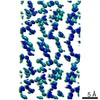
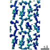
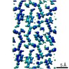
解像度: 0.67 Å / 解像度の算出法: DIFFRACTION PATTERN/LAYERLINES / 対称性のタイプ: 3D CRYSTAL | ||||||||||||||||||||||||
原子モデル構築 | プロトコル: AB INITIO MODEL / 空間: RECIPROCAL | ||||||||||||||||||||||||
精密化 | 解像度: 0.67→6.5 Å / Cor.coef. Fo:Fc: 0.904 / Cor.coef. Fo:Fc free: 0.957 / SU B: 0.523 / SU ML: 0.017 / 交差検証法: THROUGHOUT / σ(F): 1.4 / ESU R: 0.024 / ESU R Free: 0.023 詳細: HYDROGENS HAVE BEEN USED IF PRESENT IN THE INPUT U VALUES : REFINED INDIVIDUALLY
| ||||||||||||||||||||||||
溶媒の処理 | イオンプローブ半径: 0.8 Å / 減衰半径: 0.8 Å / VDWプローブ半径: 1.2 Å | ||||||||||||||||||||||||
原子変位パラメータ | Biso max: 3.82 Å2 / Biso min: 1.51 Å2
| ||||||||||||||||||||||||
精密化ステップ | サイクル: final / 解像度: 0.67→6.5 Å
| ||||||||||||||||||||||||
LS精密化 シェル | 解像度: 0.67→0.705 Å / Rfactor Rfree error: 0
|