 Movie
Movie Controller


+ Open data

- Basic information
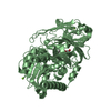
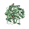
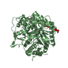
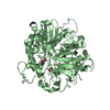
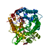
| Entry | Database: PDB / ID: 6kdc | ||||||
|---|---|---|---|---|---|---|---|
| Title | crystal structure of Fpglu1 from fervidobacterium pennivoraus | ||||||
 Components Components | Beta-glucosidase/6-phospho-beta-glucosidase/beta-galactosidase | ||||||
 Keywords Keywords | HYDROLASE / glycoside hydrolase | ||||||
| Function / homology |  Function and homology information Function and homology information | ||||||
| Biological species |  Fervidobacterium pennivorans DSM 9078 (bacteria) Fervidobacterium pennivorans DSM 9078 (bacteria) | ||||||
| Method |  X-RAY DIFFRACTION / SYNCHROTRON / MOLECULAR REPLACEMENT / Resolution: 1.85 Å X-RAY DIFFRACTION / SYNCHROTRON / MOLECULAR REPLACEMENT / Resolution: 1.85 Å | ||||||
 Authors Authors | Yu, S. / LiuQing, C. | ||||||
 Citation Citation | Journal: To Be Published Title: crystal structure of fpglu1 Authors: Yu, S. | ||||||
| History |
|
- Structure visualization
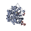
| Structure viewer | Molecule: MolmilJmol/JSmol |
|---|
- Downloads & links
-Download
| PDBx/mmCIF format | 6kdc.cif.gz | 112.8 KB | Display | PDBx/mmCIF format |
|---|---|---|---|---|
| PDB format | pdb6kdc.ent.gz | 86.6 KB | Display | PDB format |
| PDBx/mmJSON format | 6kdc.json.gz | Tree view | PDBx/mmJSON format | |
| Others |  Other downloads Other downloads |
-Validation report
| Arichive directory | https://data.pdbj.org/pub/pdb/validation_reports/kd/6kdcftp://data.pdbj.org/pub/pdb/validation_reports/kd/6kdc | HTTPS FTP |
|---|
-Related structure data
| Similar structure data |
|---|

-Links
 PDBj
PDBj
- Assembly
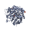
| Deposited unit | 
| ||||||||
|---|---|---|---|---|---|---|---|---|---|
| 1 |
| ||||||||
| Unit cell |
| ||||||||
| Components on special symmetry positions |
|
-Components
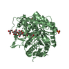
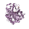
| #1: Protein | Mass: 54691.961 Da / Num. of mol.: 1 Source method: isolated from a genetically manipulated source Source: (gene. exp.) Fervidobacterium pennivorans DSM 9078 (bacteria)Strain: DSM 9078 / Ven5 / Gene: Ferpe_1843 / Production host: | ||||||
|---|---|---|---|---|---|---|---|
| #2: Chemical | ChemComp-GOL /   Mass: 92.094 Da / Num. of mol.: 5 / Source method: obtained synthetically / Formula: C3H8O3 Mass: 92.094 Da / Num. of mol.: 5 / Source method: obtained synthetically / Formula: C3H8O3#3: Chemical |   Mass: 58.933 Da / Num. of mol.: 2 / Source method: obtained synthetically / Formula: Co Mass: 58.933 Da / Num. of mol.: 2 / Source method: obtained synthetically / Formula: Co#4: Water | ChemComp-HOH / |  Mass: 18.015 Da / Num. of mol.: 293 / Source method: isolated from a natural source / Formula: H2O Mass: 18.015 Da / Num. of mol.: 293 / Source method: isolated from a natural source / Formula: H2OHas ligand of interest | N | |
-Experimental details
-Experiment
| Experiment | Method: X-RAY DIFFRACTION / Number of used crystals: 1 |
|---|
- Sample preparation
| Crystal | Density Matthews: 2.49 Å3/Da / Density % sol: 50.57 % |
|---|---|
| Crystal grow | Temperature: 291 K / Method: vapor diffusion, hanging drop / Details: PEG4000 |
-Data collection
| Diffraction | Mean temperature: 100 K / Serial crystal experiment: N |
|---|---|
| Diffraction source | Source: SYNCHROTRON / Site: SSRF  / Beamline: BL17U1 / Wavelength: 0.989 Å / Beamline: BL17U1 / Wavelength: 0.989 Å |
| Detector | Type: MAC Science DIP-320 / Detector: IMAGE PLATE / Date: Jul 1, 2016 |
| Radiation | Protocol: SINGLE WAVELENGTH / Monochromatic (M) / Laue (L): M / Scattering type: x-ray |
| Radiation wavelength | Wavelength: 0.989 Å / Relative weight: 1 |
| Reflection | Resolution: 1.85→50 Å / Num. obs: 46813 / % possible obs: 100 % / Redundancy: 9.6 % / Rpim(I) all: 0.028 / Rrim(I) all: 0.106 / Net I/σ(I): 4 |
| Reflection shell | Resolution: 1.85→1.99 Å / Redundancy: 14.4 % / Mean I/σ(I) obs: 3.1 / Num. unique obs: 2715 / CC1/2: 0.985 / Rpim(I) all: 0.132 / Rrim(I) all: 0.507 / Χ2: 1.004 |
- Processing
| Software |
| ||||||||||||||||
|---|---|---|---|---|---|---|---|---|---|---|---|---|---|---|---|---|---|
| Refinement | Method to determine structure: MOLECULAR REPLACEMENT / Resolution: 1.85→49.63 Å / Cross valid method: FREE R-VALUE
| ||||||||||||||||
| Refinement step | Cycle: LAST / Resolution: 1.85→49.63 Å
| ||||||||||||||||
| LS refinement shell | Resolution: 1.85→1.91 Å
|
