 Movie
Movie Controller


+ Open data

- Basic information
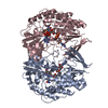
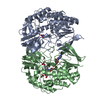
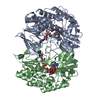
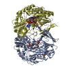
| Entry | Database: PDB / ID: 6kbw | ||||||
|---|---|---|---|---|---|---|---|
| Title | Crystal structure of Tmm from Myroides profundi D25 | ||||||
 Components Components | Trimethylamine monooxygenase | ||||||
 Keywords Keywords | FLAVOPROTEIN / flavin-containing monooxygenase | ||||||
| Function / homology |  Function and homology information Function and homology informationtrimethylamine monooxygenase / N,N-dimethylaniline monooxygenase activity / NADP binding / flavin adenine dinucleotide binding Similarity search - Function | ||||||
| Biological species |  Myroides profundi (bacteria) Myroides profundi (bacteria) | ||||||
| Method |  X-RAY DIFFRACTION / SYNCHROTRON / MOLECULAR REPLACEMENT / Resolution: 1.686 Å X-RAY DIFFRACTION / SYNCHROTRON / MOLECULAR REPLACEMENT / Resolution: 1.686 Å | ||||||
 Authors Authors | Li, C.Y. / Zhang, Y.Z. | ||||||
 Citation Citation | Journal: Sci Adv / Year: 2021 Title: Oxidation of trimethylamine to trimethylamine N-oxide facilitates high hydrostatic pressure tolerance in a generalist bacterial lineage. Authors: Qin, Q.L. / Wang, Z.B. / Su, H.N. / Chen, X.L. / Miao, J. / Wang, X.J. / Li, C.Y. / Zhang, X.Y. / Li, P.Y. / Wang, M. / Fang, J. / Lidbury, I. / Zhang, W. / Zhang, X.H. / Yang, G.P. / Chen, Y. / Zhang, Y.Z. | ||||||
| History |
|
- Structure visualization
| Structure viewer | Molecule: MolmilJmol/JSmol |
|---|
- Downloads & links
-Download
| PDBx/mmCIF format | 6kbw.cif.gz | 221.8 KB | Display | PDBx/mmCIF format |
|---|---|---|---|---|
| PDB format | pdb6kbw.ent.gz | 173.4 KB | Display | PDB format |
| PDBx/mmJSON format | 6kbw.json.gz | Tree view | PDBx/mmJSON format | |
| Others |  Other downloads Other downloads |
-Validation report
| Arichive directory | https://data.pdbj.org/pub/pdb/validation_reports/kb/6kbwftp://data.pdbj.org/pub/pdb/validation_reports/kb/6kbw | HTTPS FTP |
|---|
-Related structure data
| Related structure data | 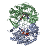 5ipyS S: Starting model for refinement |
|---|---|
| Similar structure data |


-Links
 PDBj
PDBj- Assembly
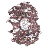
| Deposited unit | 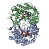
| ||||||||
|---|---|---|---|---|---|---|---|---|---|
| 1 |
| ||||||||
| Unit cell |
|
-Components
| #1: Protein | Mass: 54932.699 Da / Num. of mol.: 2 Source method: isolated from a genetically manipulated source Source: (gene. exp.) Myroides profundi (bacteria) / Gene: MPR_3295 / Production host: #2: Chemical |   Mass: 743.405 Da / Num. of mol.: 2 / Source method: obtained synthetically / Formula: C21H28N7O17P3 Mass: 743.405 Da / Num. of mol.: 2 / Source method: obtained synthetically / Formula: C21H28N7O17P3#3: Chemical |   Mass: 785.550 Da / Num. of mol.: 2 / Source method: obtained synthetically / Formula: C27H33N9O15P2 / Comment: FAD*YM Mass: 785.550 Da / Num. of mol.: 2 / Source method: obtained synthetically / Formula: C27H33N9O15P2 / Comment: FAD*YM#4: Water | ChemComp-HOH / |  Mass: 18.015 Da / Num. of mol.: 846 / Source method: isolated from a natural source / Formula: H2O Mass: 18.015 Da / Num. of mol.: 846 / Source method: isolated from a natural source / Formula: H2OHas ligand of interest | N | |
|---|
-Experimental details
-Experiment
| Experiment | Method: X-RAY DIFFRACTION / Number of used crystals: 1 |
|---|
- Sample preparation
| Crystal | Density Matthews: 2.48 Å3/Da / Density % sol: 50.32 % |
|---|---|
| Crystal grow | Temperature: 293 K / Method: vapor diffusion / pH: 6.5 Details: 0.1 M Magnesium acetate 0.1M Mes, pH 6.5 10%(w/v) PEG 10000 |
-Data collection
| Diffraction | Mean temperature: 100 K / Serial crystal experiment: N |
|---|---|
| Diffraction source | Source: SYNCHROTRON / Site: SSRF  / Beamline: BL17U1 / Wavelength: 0.987 Å / Beamline: BL17U1 / Wavelength: 0.987 Å |
| Detector | Type: DECTRIS EIGER X 16M / Detector: PIXEL / Date: Apr 2, 2018 |
| Radiation | Protocol: SINGLE WAVELENGTH / Monochromatic (M) / Laue (L): M / Scattering type: x-ray |
| Radiation wavelength | Wavelength: 0.987 Å / Relative weight: 1 |
| Reflection | Resolution: 1.686→81.96 Å / Num. obs: 115098 / % possible obs: 93.4 % / Redundancy: 11.3 % / Rmerge(I) obs: 0.082 / Rpim(I) all: 0.034 / Rrim(I) all: 0.089 / Net I/σ(I): 17.8 |
| Reflection shell | Resolution: 1.69→1.78 Å / Redundancy: 5.5 % / Rmerge(I) obs: 0.44 / Num. unique obs: 12333 / Rpim(I) all: 0.317 / % possible all: 69.9 |
- Processing
| Software |
| ||||||||||||||||||||||||||||||||||||||||||||||||||||||||||||||||||
|---|---|---|---|---|---|---|---|---|---|---|---|---|---|---|---|---|---|---|---|---|---|---|---|---|---|---|---|---|---|---|---|---|---|---|---|---|---|---|---|---|---|---|---|---|---|---|---|---|---|---|---|---|---|---|---|---|---|---|---|---|---|---|---|---|---|---|---|
| Refinement | Method to determine structure: MOLECULAR REPLACEMENT Starting model: 5IPY Resolution: 1.686→81.956 Å / SU ML: 0.17 / Cross valid method: THROUGHOUT / σ(F): 0 / Phase error: 21.25
| ||||||||||||||||||||||||||||||||||||||||||||||||||||||||||||||||||
| Solvent computation | Shrinkage radii: 0.95 Å / VDW probe radii: 1.2 Å / Bsol: 39.087 Å2 / ksol: 0.335 e/Å3 | ||||||||||||||||||||||||||||||||||||||||||||||||||||||||||||||||||
| Displacement parameters | Biso max: 65.14 Å2 / Biso mean: 27.91 Å2 / Biso min: 10.69 Å2
| ||||||||||||||||||||||||||||||||||||||||||||||||||||||||||||||||||
| Refinement step | Cycle: final / Resolution: 1.686→81.956 Å
| ||||||||||||||||||||||||||||||||||||||||||||||||||||||||||||||||||
| Refine LS restraints |
| ||||||||||||||||||||||||||||||||||||||||||||||||||||||||||||||||||
| LS refinement shell | Refine-ID: X-RAY DIFFRACTION / Rfactor Rfree error: 0
|
