 Movie
Movie Controller


+ Open data

- Basic information
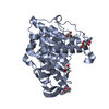
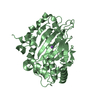
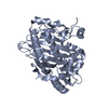
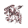
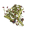
| Entry | Database: PDB / ID: 6kaw | ||||||
|---|---|---|---|---|---|---|---|
| Title | Crystal structure of CghA | ||||||
 Components Components | CghA | ||||||
 Keywords Keywords | UNKNOWN FUNCTION / Diels-Alderase | ||||||
| Function / homology | : / : / Diels-Alderase-like / : / Isomerases; Intramolecular lyases / isomerase activity / Diels-Alderase cghA Function and homology information Function and homology information | ||||||
| Biological species |  Chaetomium globosum (fungus) Chaetomium globosum (fungus) | ||||||
| Method |  X-RAY DIFFRACTION / SYNCHROTRON / SAD / Resolution: 2.01 Å X-RAY DIFFRACTION / SYNCHROTRON / SAD / Resolution: 2.01 Å | ||||||
 Authors Authors | Hara, K. / Hashimoto, H. / Yokoyama, M. / Sato, M. / Watanabe, K. | ||||||
 Citation Citation | Journal: Nat Catal / Year: 2021 Title: Catalytic mechanism and endo-to-exo selectivity reversion of an octalin-forming natural Diels-Alderase Authors: Sato, M. / Kishimoto, S. / Yokoyama, M. / Jamieson, C.S. / Narita, K. / Maeda, N. / Hara, K. / Hashimoto, H. / Tsunematsu, Y. / Houk, K.N. / Tang, Y. / Watanabe, K. | ||||||
| History |
|
- Structure visualization
| Structure viewer | Molecule: MolmilJmol/JSmol |
|---|
- Downloads & links
-Download
| PDBx/mmCIF format | 6kaw.cif.gz | 96.7 KB | Display | PDBx/mmCIF format |
|---|---|---|---|---|
| PDB format | pdb6kaw.ent.gz | 71.3 KB | Display | PDB format |
| PDBx/mmJSON format | 6kaw.json.gz | Tree view | PDBx/mmJSON format | |
| Others |  Other downloads Other downloads |
-Validation report
| Arichive directory | https://data.pdbj.org/pub/pdb/validation_reports/ka/6kawftp://data.pdbj.org/pub/pdb/validation_reports/ka/6kaw | HTTPS FTP |
|---|
-Related structure data


-Links
 PDBj
PDBj- Assembly
| Deposited unit | 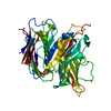
| ||||||||
|---|---|---|---|---|---|---|---|---|---|
| 1 |
| ||||||||
| Unit cell |
|
-Components
| #1: Protein | Mass: 44164.992 Da / Num. of mol.: 1 Source method: isolated from a genetically manipulated source Source: (gene. exp.) Chaetomium globosum (strain ATCC 6205 / CBS 148.51 / DSM 1962 / NBRC 6347 / NRRL 1970) (fungus)Strain: ATCC 6205 / CBS 148.51 / DSM 1962 / NBRC 6347 / NRRL 1970 Gene: CHGG_02368 / Production host:  |
|---|---|
| #2: Water | ChemComp-HOH /  Mass: 18.015 Da / Num. of mol.: 417 / Source method: isolated from a natural source / Formula: H2O Mass: 18.015 Da / Num. of mol.: 417 / Source method: isolated from a natural source / Formula: H2O |
-Experimental details
-Experiment
| Experiment | Method: X-RAY DIFFRACTION / Number of used crystals: 1 |
|---|
- Sample preparation
| Crystal | Density Matthews: 3.11 Å3/Da / Density % sol: 60.48 % |
|---|---|
| Crystal grow | Temperature: 293 K / Method: vapor diffusion, hanging drop / Details: PEG 4000, MES |
-Data collection
| Diffraction | Mean temperature: 100 K / Serial crystal experiment: N |
|---|---|
| Diffraction source | Source: SYNCHROTRON / Site: Photon Factory  / Beamline: BL-17A / Wavelength: 0.98 Å / Beamline: BL-17A / Wavelength: 0.98 Å |
| Detector | Type: DECTRIS PILATUS3 S 6M / Detector: PIXEL / Date: Dec 14, 2016 |
| Radiation | Protocol: SINGLE WAVELENGTH / Monochromatic (M) / Laue (L): M / Scattering type: x-ray |
| Radiation wavelength | Wavelength: 0.98 Å / Relative weight: 1 |
| Reflection | Resolution: 2.01→20 Å / Num. obs: 38207 / % possible obs: 99.8 % / Redundancy: 13 % / Rmerge(I) obs: 0.181 / Net I/σ(I): 10.4 |
| Reflection shell | Resolution: 2.01→2.13 Å / Rmerge(I) obs: 0.864 / Num. unique obs: 5995 |
- Processing
| Software |
| ||||||||||||||||||||||||||||||||||||||||||||||||||||||||||||||||||||||||||||||||||||||||||||||||||
|---|---|---|---|---|---|---|---|---|---|---|---|---|---|---|---|---|---|---|---|---|---|---|---|---|---|---|---|---|---|---|---|---|---|---|---|---|---|---|---|---|---|---|---|---|---|---|---|---|---|---|---|---|---|---|---|---|---|---|---|---|---|---|---|---|---|---|---|---|---|---|---|---|---|---|---|---|---|---|---|---|---|---|---|---|---|---|---|---|---|---|---|---|---|---|---|---|---|---|---|
| Refinement | Method to determine structure: SAD / Resolution: 2.01→20 Å / SU ML: 0.18 / Cross valid method: FREE R-VALUE / σ(F): 1.34 / Phase error: 21.66
| ||||||||||||||||||||||||||||||||||||||||||||||||||||||||||||||||||||||||||||||||||||||||||||||||||
| Solvent computation | Shrinkage radii: 0.9 Å / VDW probe radii: 1.11 Å | ||||||||||||||||||||||||||||||||||||||||||||||||||||||||||||||||||||||||||||||||||||||||||||||||||
| Refinement step | Cycle: LAST / Resolution: 2.01→20 Å
| ||||||||||||||||||||||||||||||||||||||||||||||||||||||||||||||||||||||||||||||||||||||||||||||||||
| Refine LS restraints |
| ||||||||||||||||||||||||||||||||||||||||||||||||||||||||||||||||||||||||||||||||||||||||||||||||||
| LS refinement shell |
|
