 Movie
Movie Controller


+ Open data

- Basic information
| Entry | Database: PDB / ID: 6ka4 | ||||||
|---|---|---|---|---|---|---|---|
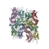
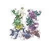
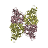
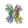
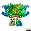
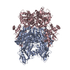
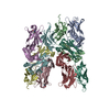
| Title | Cryo-EM structure of the AtMLKL3 tetramer | ||||||
 Components Components | F22L4.1 protein | ||||||
 Keywords Keywords | MEMBRANE PROTEIN / Plant MLKLs / Cryo-EM / Tetramer | ||||||
| Function / homology |  Function and homology information Function and homology information | ||||||
| Biological species |  | ||||||
| Method | ELECTRON MICROSCOPY / single particle reconstruction / cryo EM / Resolution: 3.4 Å | ||||||
 Authors Authors | Lisa, M. / Huang, M. / Zhang, X. / Ryohei, T.N. / Leila, B.K. / Isabel, M.L.S. / Florence, J. / Viera, K. / Dmitry, L. / Jane, E.P. ...Lisa, M. / Huang, M. / Zhang, X. / Ryohei, T.N. / Leila, B.K. / Isabel, M.L.S. / Florence, J. / Viera, K. / Dmitry, L. / Jane, E.P. / James, M.M. / Kay, H. / Paul, S.L. / Chai, J. / Takaki, M. | ||||||
 Citation Citation | Journal: To Be Published Title: Cryo-EM structure of the AtMLKL3 tetramer Authors: Lisa, M. / Huang, M. / Zhang, X. / Ryohei, T.N. / Leila, B.K. / Isabel, M.L.S. / Florence, J. / Viera, K. / Dmitry, L. / Jane, E.P. / James, M.M. / Kay, H. / Paul, S.L. / Chai, J. / Takaki, M. | ||||||
| History |
|
- Structure visualization
| Movie |
Movie viewer |
|---|---|
| Structure viewer | Molecule: MolmilJmol/JSmol |
- Downloads & links
-Download
| PDBx/mmCIF format | 6ka4.cif.gz | 383.7 KB | Display | PDBx/mmCIF format |
|---|---|---|---|---|
| PDB format | pdb6ka4.ent.gz | 313.6 KB | Display | PDB format |
| PDBx/mmJSON format | 6ka4.json.gz | Tree view | PDBx/mmJSON format | |
| Others |  Other downloads Other downloads |
-Validation report
| Arichive directory | https://data.pdbj.org/pub/pdb/validation_reports/ka/6ka4ftp://data.pdbj.org/pub/pdb/validation_reports/ka/6ka4 | HTTPS FTP |
|---|
-Related structure data
| Related structure data |  9954MC 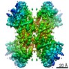 0868C  6lbaC M: map data used to model this data C: citing same article ( |
|---|---|
| Similar structure data |
-Links
 PDBj
PDBj- Assembly
| Deposited unit | 
|
|---|---|
| 1 |
|
-Components
| #1: Protein | Mass: 80046.188 Da / Num. of mol.: 4 Source method: isolated from a genetically manipulated source Source: (gene. exp.) Production host:  Spodoptera aff. frugiperda 1 BOLD-2017 (butterflies/moths) Spodoptera aff. frugiperda 1 BOLD-2017 (butterflies/moths)References: UniProt: Q9LMN2 |
|---|
-Experimental details
-Experiment
| Experiment | Method: ELECTRON MICROSCOPY |
|---|---|
| EM experiment | Aggregation state: PARTICLE / 3D reconstruction method: single particle reconstruction |
- Sample preparation
| Component | Name: Mixed lineage kinase domain-like (MLKL) protein / Type: COMPLEX / Entity ID: all / Source: RECOMBINANT |
|---|---|
| Source (natural) | Organism: |
| Source (recombinant) | Organism: Spodoptera aff. frugiperda 1 BOLD-2017 (butterflies/moths) |
| Buffer solution | pH: 8 |
| Specimen | Embedding applied: NO / Shadowing applied: NO / Staining applied: NO / Vitrification applied: YES |
| Vitrification | Cryogen name: ETHANE |
- Electron microscopy imaging
| Experimental equipment |  Model: Titan Krios / Image courtesy: FEI Company |
|---|---|
| Microscopy | Model: FEI TITAN KRIOS |
| Electron gun | Electron source:  FIELD EMISSION GUN / Accelerating voltage: 300 kV / Illumination mode: FLOOD BEAM FIELD EMISSION GUN / Accelerating voltage: 300 kV / Illumination mode: FLOOD BEAM |
| Electron lens | Mode: BRIGHT FIELD |
| Image recording | Electron dose: 1.5625 e/Å2 / Film or detector model: GATAN K2 SUMMIT (4k x 4k) |
- Processing
| Software | Name: PHENIX / Version: 1.18.2_3874: / Classification: refinement | ||||||||||||||||||||||||
|---|---|---|---|---|---|---|---|---|---|---|---|---|---|---|---|---|---|---|---|---|---|---|---|---|---|
| CTF correction | Type: NONE | ||||||||||||||||||||||||
| 3D reconstruction | Resolution: 3.4 Å / Resolution method: FSC 0.143 CUT-OFF / Num. of particles: 983779 / Symmetry type: POINT | ||||||||||||||||||||||||
| Refine LS restraints |
|
