 Movie
Movie Controller

[English] 日本語
 Yorodumi
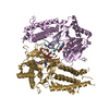
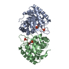
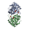
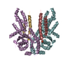
- PDB-6k8w: Crystal structure of N-domain with NADP of baterial malonyl-CoA r... -
+ Open data

- Basic information
| Entry | Database: PDB / ID: 6k8w | ||||||
|---|---|---|---|---|---|---|---|
| Title | Crystal structure of N-domain with NADP of baterial malonyl-CoA reductase | ||||||
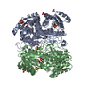
 Components Components | NAD-dependent epimerase/dehydratase:Short-chain dehydrogenase/reductase SDR | ||||||
 Keywords Keywords | OXIDOREDUCTASE / SDR domain | ||||||
| Function / homology | fatty acid elongation / oxidoreductase activity, acting on the CH-OH group of donors, NAD or NADP as acceptor / short chain dehydrogenase / Short-chain dehydrogenase/reductase SDR / NAD(P)-binding domain superfamily / nucleotide binding / NADP NICOTINAMIDE-ADENINE-DINUCLEOTIDE PHOSPHATE / NAD-dependent epimerase/dehydratase:Short-chain dehydrogenase/reductase SDR Function and homology information Function and homology information | ||||||
| Biological species |  Porphyrobacter dokdonensis DSW-74 (bacteria) Porphyrobacter dokdonensis DSW-74 (bacteria) | ||||||
| Method |  X-RAY DIFFRACTION / SYNCHROTRON / MOLECULAR REPLACEMENT / Resolution: 3.17 Å X-RAY DIFFRACTION / SYNCHROTRON / MOLECULAR REPLACEMENT / Resolution: 3.17 Å | ||||||
 Authors Authors | Kim, S. / Kim, K.-J. | ||||||
 Citation Citation | Journal: Environ.Microbiol. / Year: 2020 Title: Structural insight into bi-functional malonyl-CoA reductase. Authors: Son, H.F. / Kim, S. / Seo, H. / Hong, J. / Lee, D. / Jin, K.S. / Park, S. / Kim, K.J. | ||||||
| History |
|
- Structure visualization
| Structure viewer | Molecule: MolmilJmol/JSmol |
|---|
- Downloads & links
-Download
| PDBx/mmCIF format | 6k8w.cif.gz | 213.2 KB | Display | PDBx/mmCIF format |
|---|---|---|---|---|
| PDB format | pdb6k8w.ent.gz | 169.5 KB | Display | PDB format |
| PDBx/mmJSON format | 6k8w.json.gz | Tree view | PDBx/mmJSON format | |
| Others |  Other downloads Other downloads |
-Validation report
| Arichive directory | https://data.pdbj.org/pub/pdb/validation_reports/k8/6k8wftp://data.pdbj.org/pub/pdb/validation_reports/k8/6k8w | HTTPS FTP |
|---|
-Related structure data
| Related structure data | 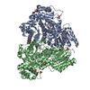 6k8sC 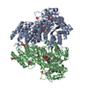 6k8tC  6k8uC  6k8vSC S: Starting model for refinement C: citing same article ( |
|---|---|
| Similar structure data |


-Links
 PDBj
PDBj

- Assembly
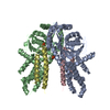
| Deposited unit | 
| ||||||||
|---|---|---|---|---|---|---|---|---|---|
| 1 |
| ||||||||
| Unit cell |
|
-Components
| #1: Protein | Mass: 61269.301 Da / Num. of mol.: 2 / Fragment: SDR N-domain Source method: isolated from a genetically manipulated source Source: (gene. exp.) Porphyrobacter dokdonensis DSW-74 (bacteria)Gene: I603_0811 / Plasmid: pET30a / Production host: #2: Chemical |   Mass: 743.405 Da / Num. of mol.: 2 / Source method: obtained synthetically / Formula: C21H28N7O17P3 / Feature type: SUBJECT OF INVESTIGATION Mass: 743.405 Da / Num. of mol.: 2 / Source method: obtained synthetically / Formula: C21H28N7O17P3 / Feature type: SUBJECT OF INVESTIGATION#3: Chemical | ChemComp-SO4 / |   Mass: 96.063 Da / Num. of mol.: 1 / Source method: obtained synthetically / Formula: SO4 / Feature type: SUBJECT OF INVESTIGATION Mass: 96.063 Da / Num. of mol.: 1 / Source method: obtained synthetically / Formula: SO4 / Feature type: SUBJECT OF INVESTIGATION#4: Water | ChemComp-HOH / |  Mass: 18.015 Da / Num. of mol.: 25 / Source method: isolated from a natural source / Formula: H2O Mass: 18.015 Da / Num. of mol.: 25 / Source method: isolated from a natural source / Formula: H2OHas ligand of interest | Y | |
|---|
-Experimental details
-Experiment
| Experiment | Method: X-RAY DIFFRACTION / Number of used crystals: 1 |
|---|
- Sample preparation
| Crystal | Density Matthews: 2.5 Å3/Da / Density % sol: 50.85 % |
|---|---|
| Crystal grow | Temperature: 295 K / Method: vapor diffusion, hanging drop / pH: 8 / Details: PEG3350, Li2SO4, Tris |
-Data collection
| Diffraction | Mean temperature: 100 K / Serial crystal experiment: N |
|---|---|
| Diffraction source | Source: SYNCHROTRON / Site: PAL/PLS  / Beamline: 7A (6B, 6C1) / Wavelength: 0.97933 Å / Beamline: 7A (6B, 6C1) / Wavelength: 0.97933 Å |
| Detector | Type: ADSC QUANTUM 270 / Detector: CCD / Date: Dec 18, 2018 |
| Radiation | Protocol: SINGLE WAVELENGTH / Monochromatic (M) / Laue (L): M / Scattering type: x-ray |
| Radiation wavelength | Wavelength: 0.97933 Å / Relative weight: 1 |
| Reflection | Resolution: 3.17→50 Å / Num. obs: 20735 / % possible obs: 97.4 % / Redundancy: 4.6 % / CC1/2: 0.979 / Rmerge(I) obs: 0.153 / Rpim(I) all: 0.074 / Rsym value: 0.171 / Net I/σ(I): 13.1 |
| Reflection shell | Resolution: 3.2→3.26 Å / Rmerge(I) obs: 0.302 / Num. unique obs: 987 / CC1/2: 0.845 / Rpim(I) all: 0.173 / Rsym value: 0.351 |
- Processing
| Software |
| ||||||||||||||||||||||||||||||||||||||||||||||||||||||||||||
|---|---|---|---|---|---|---|---|---|---|---|---|---|---|---|---|---|---|---|---|---|---|---|---|---|---|---|---|---|---|---|---|---|---|---|---|---|---|---|---|---|---|---|---|---|---|---|---|---|---|---|---|---|---|---|---|---|---|---|---|---|---|
| Refinement | Method to determine structure: MOLECULAR REPLACEMENT Starting model: 6K8V Resolution: 3.17→33.76 Å / Cor.coef. Fo:Fc: 0.916 / Cor.coef. Fo:Fc free: 0.845 / SU B: 31.898 / SU ML: 0.513 / Cross valid method: THROUGHOUT / σ(F): 0 / ESU R Free: 0.592 Details: HYDROGENS HAVE BEEN ADDED IN THE RIDING POSITIONS U VALUES : REFINED INDIVIDUALLY
| ||||||||||||||||||||||||||||||||||||||||||||||||||||||||||||
| Solvent computation | Ion probe radii: 0.8 Å / Shrinkage radii: 0.8 Å / VDW probe radii: 1.2 Å | ||||||||||||||||||||||||||||||||||||||||||||||||||||||||||||
| Displacement parameters | Biso max: 138.07 Å2 / Biso mean: 43.266 Å2 / Biso min: 0.5 Å2
| ||||||||||||||||||||||||||||||||||||||||||||||||||||||||||||
| Refinement step | Cycle: final / Resolution: 3.17→33.76 Å
| ||||||||||||||||||||||||||||||||||||||||||||||||||||||||||||
| Refine LS restraints |
| ||||||||||||||||||||||||||||||||||||||||||||||||||||||||||||
| LS refinement shell | Resolution: 3.174→3.256 Å / Rfactor Rfree error: 0 / Total num. of bins used: 20
|
