[English] 日本語
Yorodumi
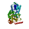
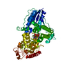
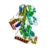
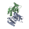
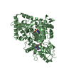
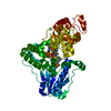
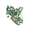
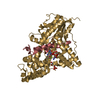
- PDB-6k8k: Crystal structure of Arabidopsis thaliana BIC2-CRY2 complex -

+
Open data


ID or keywords:

Loading...

-
Basic information

Entry
Database: PDB / ID: 6k8k
TitleCrystal structure of Arabidopsis thaliana BIC2-CRY2 complex
Components
  • Cryptochrome-2
  • Protein BIC2
KeywordsSIGNALING PROTEIN / Cryptochromes / BICs / inactivation
Function / homology
Function and homology information


flavin adenine dinucleotide metabolic process / long-day photoperiodism, flowering / response to absence of light / circadian regulation of calcium ion oscillation / response to strigolactone / regulation of meristem growth / response to low fluence blue light stimulus by blue low-fluence system / response to karrikin / regulation of leaf morphogenesis / regulation of photoperiodism, flowering ...flavin adenine dinucleotide metabolic process / long-day photoperiodism, flowering / response to absence of light / circadian regulation of calcium ion oscillation / response to strigolactone / regulation of meristem growth / response to low fluence blue light stimulus by blue low-fluence system / response to karrikin / regulation of leaf morphogenesis / regulation of photoperiodism, flowering / blue light signaling pathway / regulation of flower development / positive regulation of flower development / phototropism / stomatal movement / response to blue light / blue light photoreceptor activity / response to water deprivation / plant-type vacuole / response to light stimulus / FAD binding / regulation of circadian rhythm / PML body / circadian rhythm / positive regulation of reactive oxygen species metabolic process / chromatin organization / defense response to virus / nuclear body / chromatin remodeling / protein homodimerization activity / ATP binding / identical protein binding / metal ion binding / nucleus / cytoplasm
Similarity search - Function
Protein BIC / Cryptochrome, plant / DNA photolyases class 1 signature 2. / Cryptochrome/DNA photolyase class 1, conserved site, C-terminal / DNA photolyases class 1 signature 1. / DNA Cyclobutane Dipyrimidine Photolyase, subunit A; domain 3 / DNA Cyclobutane Dipyrimidine Photolyase, subunit A, domain 3 / Serine Threonine Protein Phosphatase 5, Tetratricopeptide repeat - #80 / Cryptochrome/DNA photolyase class 1 / Cryptochrome/DNA photolyase, FAD-binding domain ...Protein BIC / Cryptochrome, plant / DNA photolyases class 1 signature 2. / Cryptochrome/DNA photolyase class 1, conserved site, C-terminal / DNA photolyases class 1 signature 1. / DNA Cyclobutane Dipyrimidine Photolyase, subunit A; domain 3 / DNA Cyclobutane Dipyrimidine Photolyase, subunit A, domain 3 / Serine Threonine Protein Phosphatase 5, Tetratricopeptide repeat - #80 / Cryptochrome/DNA photolyase class 1 / Cryptochrome/DNA photolyase, FAD-binding domain / FAD binding domain of DNA photolyase / DNA photolyase, N-terminal / Cryptochrome/photolyase, N-terminal domain superfamily / DNA photolyase / Photolyase/cryptochrome alpha/beta domain profile. / Cryptochrome/DNA photolyase, FAD-binding domain-like superfamily / HUPs / Rossmann-like alpha/beta/alpha sandwich fold / Serine Threonine Protein Phosphatase 5, Tetratricopeptide repeat / Alpha Horseshoe / Rossmann fold / Orthogonal Bundle / 3-Layer(aba) Sandwich / Mainly Alpha / Alpha Beta
Similarity search - Domain/homology
ADENOSINE MONOPHOSPHATE / FLAVIN-ADENINE DINUCLEOTIDE / Cryptochrome-2 / Protein BIC2
Similarity search - Component
Biological speciesArabidopsis thaliana (thale cress)
MethodX-RAY DIFFRACTION / SYNCHROTRON / MOLECULAR REPLACEMENT / Resolution: 2.5 Å
AuthorsWang, X. / Ma, L. / Guan, Z. / Yin, P.
CitationJournal: Nat.Struct.Mol.Biol. / Year: 2020
Title: Structural insights into BIC-mediated inactivation of Arabidopsis cryptochrome 2.
Authors: Ma, L. / Wang, X. / Guan, Z. / Wang, L. / Wang, Y. / Zheng, L. / Gong, Z. / Shen, C. / Wang, J. / Zhang, D. / Liu, Z. / Yin, P.
History
DepositionJun 12, 2019Deposition site: PDBJ / Processing site: PDBJ
Revision 1.0May 13, 2020Provider: repository / Type: Initial release
Revision 1.1May 27, 2020Group: Database references / Category: citation / citation_author
Item: _citation.journal_volume / _citation.page_first ..._citation.journal_volume / _citation.page_first / _citation.page_last / _citation.pdbx_database_id_PubMed / _citation.title / _citation_author.identifier_ORCID
Revision 1.2Nov 22, 2023Group: Data collection / Database references ...Data collection / Database references / Derived calculations / Refinement description
Category: chem_comp_atom / chem_comp_bond ...chem_comp_atom / chem_comp_bond / database_2 / pdbx_initial_refinement_model / struct_conn
Item: _database_2.pdbx_DOI / _database_2.pdbx_database_accession ..._database_2.pdbx_DOI / _database_2.pdbx_database_accession / _struct_conn.pdbx_dist_value / _struct_conn.pdbx_leaving_atom_flag / _struct_conn.ptnr1_auth_asym_id / _struct_conn.ptnr1_auth_comp_id / _struct_conn.ptnr1_auth_seq_id / _struct_conn.ptnr1_label_asym_id / _struct_conn.ptnr1_label_atom_id / _struct_conn.ptnr1_label_comp_id / _struct_conn.ptnr1_label_seq_id / _struct_conn.ptnr2_auth_asym_id / _struct_conn.ptnr2_auth_seq_id / _struct_conn.ptnr2_label_asym_id

-
Structure visualization

Structure viewerMolecule:
MolmilJmol/JSmol

Downloads & links

-
Assembly

Deposited unit
A: Cryptochrome-2
E: Protein BIC2
B: Cryptochrome-2
C: Protein BIC2
D: Cryptochrome-2
F: Protein BIC2
G: Cryptochrome-2
H: Protein BIC2
hetero molecules


Theoretical massNumber of molelcules
Total (without water)314,62820
Polymers309,9998
Non-polymers4,62812
Water2,036113
1
A: Cryptochrome-2
E: Protein BIC2
hetero molecules


Theoretical massNumber of molelcules
Total (without water)78,6575
Polymers77,5002
Non-polymers1,1573
Water362
TypeNameSymmetry operationNumber
identity operation1_555x,y,z1
Buried area6320 Å2
ΔGint-48 kcal/mol
Surface area23140 Å2
MethodPISA
2
B: Cryptochrome-2
C: Protein BIC2
hetero molecules


Theoretical massNumber of molelcules
Total (without water)78,6334
Polymers77,5002
Non-polymers1,1332
Water362
TypeNameSymmetry operationNumber
identity operation1_555x,y,z1
Buried area6030 Å2
ΔGint-36 kcal/mol
Surface area22520 Å2
MethodPISA
3
D: Cryptochrome-2
F: Protein BIC2
hetero molecules


Theoretical massNumber of molelcules
Total (without water)78,6816
Polymers77,5002
Non-polymers1,1814
Water362
TypeNameSymmetry operationNumber
identity operation1_555x,y,z1
Buried area6870 Å2
ΔGint-58 kcal/mol
Surface area22530 Å2
MethodPISA
4
G: Cryptochrome-2
H: Protein BIC2
hetero molecules


Theoretical massNumber of molelcules
Total (without water)78,6575
Polymers77,5002
Non-polymers1,1573
Water362
TypeNameSymmetry operationNumber
identity operation1_555x,y,z1
Buried area6600 Å2
ΔGint-49 kcal/mol
Surface area23000 Å2
MethodPISA
Unit cell
Length a, b, c (Å)142.552, 142.552, 526.434
Angle α, β, γ (deg.)90.00, 90.00, 120.00
Int Tables number179
Space group name H-MP6522

-
Components

-
Protein , 2 types, 8 molecules ABDGECFH

#1: Protein
Cryptochrome-2 / / Atcry2 / Blue light photoreceptor / Protein PHR homolog 1 / AtPHH1 / Protein SUPPRESSOR OF elf3 20


Mass: 69579.930 Da / Num. of mol.: 4
Source method: isolated from a genetically manipulated source
Source: (gene. exp.) Arabidopsis thaliana (thale cress) / Gene: CRY2, PHH1, SEL20, At1g04400, F19P19.14 / Production host: Escherichia coli BL21(DE3) (bacteria) / Strain (production host): BL21(DE3) / References: UniProt: Q96524
#2: Protein
Protein BIC2 / BLUE-LIGHT INHIBITOR OF CRYPTOCHROMES 2


Mass: 7919.887 Da / Num. of mol.: 4
Source method: isolated from a genetically manipulated source
Source: (gene. exp.) Arabidopsis thaliana (thale cress) / Gene: BIC2, At3g44450, T22K7.130 / Production host: Escherichia coli BL21(DE3) (bacteria) / Strain (production host): BL21(DE3) / References: UniProt: Q9M280

-
Non-polymers , 4 types, 125 molecules

#3: Chemical
ChemComp-FAD / FLAVIN-ADENINE DINUCLEOTIDE / Flavin adenine dinucleotide


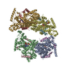
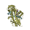
Mass: 785.550 Da / Num. of mol.: 4 / Source method: obtained synthetically / Formula: C27H33N9O15P2 / Feature type: SUBJECT OF INVESTIGATION / Comment: FAD*YM
#4: Chemical
ChemComp-AMP / ADENOSINE MONOPHOSPHATE / Adenosine monophosphate


Mass: 347.221 Da / Num. of mol.: 4 / Source method: obtained synthetically / Formula: C10H14N5O7P / Feature type: SUBJECT OF INVESTIGATION / Comment: AMP*YM
#5: Chemical
ChemComp-MG / MAGNESIUM ION


Mass: 24.305 Da / Num. of mol.: 4 / Source method: obtained synthetically / Formula: Mg / Feature type: SUBJECT OF INVESTIGATION
#6: Water ChemComp-HOH / water / Water


Mass: 18.015 Da / Num. of mol.: 113 / Source method: isolated from a natural source / Formula: H2O

-
Details

Has ligand of interestY
Sequence detailsThese conflicts are derived from GenBank AAB04996.

-
Experimental details

-
Experiment

ExperimentMethod: X-RAY DIFFRACTION / Number of used crystals: 1

-
Sample preparation

CrystalDensity Matthews: 2.49 Å3/Da / Density % sol: 50.61 %
Crystal growTemperature: 291.15 K / Method: vapor diffusion, hanging drop / Details: Tris, Calcium acetate hydrate, PEG 3350

-
Data collection

DiffractionMean temperature: 100 K / Serial crystal experiment: N
Diffraction sourceSource: SYNCHROTRON / Site: SSRF / Beamline: BL17U1 / Wavelength: 0.97919 Å
DetectorType: DECTRIS EIGER X 16M / Detector: PIXEL / Date: Mar 26, 2018
RadiationProtocol: SINGLE WAVELENGTH / Monochromatic (M) / Laue (L): M / Scattering type: x-ray
Radiation wavelengthWavelength: 0.97919 Å / Relative weight: 1
ReflectionResolution: 2.5→50 Å / Num. obs: 109254 / % possible obs: 99.2 % / Redundancy: 19.2 % / Biso Wilson estimate: 45.2 Å2 / CC1/2: 0.999 / Rmerge(I) obs: 0.101 / Rpim(I) all: 0.024 / Rrim(I) all: 0.104 / Net I/σ(I): 21.3
Reflection shellResolution: 2.5→2.54 Å / Rmerge(I) obs: 0.784 / Mean I/σ(I) obs: 4.4 / Num. unique obs: 5043 / CC1/2: 0.92 / Rpim(I) all: 0.186 / Rrim(I) all: 0.807 / % possible all: 94.6

-
Processing

Software
NameVersionClassification
PHENIX(1.15.2_3472: ???)refinement
XDSdata reduction
Aimlessdata scaling
PHASERphasing
RefinementMethod to determine structure: MOLECULAR REPLACEMENT
Starting model: 1U3C
Resolution: 2.5→48.425 Å / SU ML: 0.3 / Cross valid method: FREE R-VALUE / σ(F): 1.34 / Phase error: 24.65 / Stereochemistry target values: ML
RfactorNum. reflection% reflection
Rfree0.2232 5437 4.98 %
Rwork0.1706 --
obs0.1733 109130 99.01 %
Solvent computationShrinkage radii: 0.9 Å / VDW probe radii: 1.11 Å / Solvent model: FLAT BULK SOLVENT MODEL
Refinement stepCycle: LAST / Resolution: 2.5→48.425 Å
ProteinNucleic acidLigandSolventTotal
Num. atoms17459 0 308 113 17880
Refine LS restraints
Refine-IDTypeDev idealNumber
X-RAY DIFFRACTIONf_bond_d0.00818292
X-RAY DIFFRACTIONf_angle_d0.98924882
X-RAY DIFFRACTIONf_dihedral_angle_d20.92210699
X-RAY DIFFRACTIONf_chiral_restr0.0532631
X-RAY DIFFRACTIONf_plane_restr0.0063093
LS refinement shell
Resolution (Å)Rfactor RfreeNum. reflection RfreeRfactor RworkNum. reflection RworkRefine-ID% reflection obs (%)
2.4998-2.52820.32761760.25743157X-RAY DIFFRACTION93
2.5282-2.55790.28871730.22033300X-RAY DIFFRACTION97
2.5579-2.58910.28261900.21343353X-RAY DIFFRACTION97
2.5891-2.62190.30591820.21593351X-RAY DIFFRACTION98
2.6219-2.65640.30381790.21663418X-RAY DIFFRACTION99
2.6564-2.69270.31091750.2183422X-RAY DIFFRACTION100
2.6927-2.73120.2911610.22573419X-RAY DIFFRACTION99
2.7312-2.7720.3051750.20963426X-RAY DIFFRACTION100
2.772-2.81530.26791780.19983422X-RAY DIFFRACTION100
2.8153-2.86140.2482020.20293402X-RAY DIFFRACTION100
2.8614-2.91080.28861860.20323440X-RAY DIFFRACTION100
2.9108-2.96370.3181890.22793426X-RAY DIFFRACTION100
2.9637-3.02070.32462030.22953407X-RAY DIFFRACTION100
3.0207-3.08230.28781930.23563434X-RAY DIFFRACTION100
3.0823-3.14930.32791940.22613409X-RAY DIFFRACTION100
3.1493-3.22260.30411940.22293458X-RAY DIFFRACTION100
3.2226-3.30320.27691700.20873352X-RAY DIFFRACTION96
3.3032-3.39250.2561730.19313488X-RAY DIFFRACTION100
3.3925-3.49230.231700.18433458X-RAY DIFFRACTION100
3.4923-3.60490.21461640.173480X-RAY DIFFRACTION100
3.6049-3.73370.2031780.15973513X-RAY DIFFRACTION100
3.7337-3.88320.19151730.14993485X-RAY DIFFRACTION100
3.8832-4.05980.19251710.13993533X-RAY DIFFRACTION100
4.0598-4.27370.18121850.13963511X-RAY DIFFRACTION100
4.2737-4.54130.1882020.13373509X-RAY DIFFRACTION100
4.5413-4.89160.16711710.13193459X-RAY DIFFRACTION97
4.8916-5.38330.18091770.14343565X-RAY DIFFRACTION100
5.3833-6.1610.20641690.15793626X-RAY DIFFRACTION100
6.161-7.7570.19971920.1623670X-RAY DIFFRACTION100
7.757-48.43360.17361920.14493800X-RAY DIFFRACTION97
Refinement TLS params.Method: refined / Origin x: 55.5794 Å / Origin y: -12.9522 Å / Origin z: -67.4519 Å
111213212223313233
T0.4104 Å2-0.0391 Å2-0.0138 Å2-0.2542 Å20.0265 Å2--0.4159 Å2
L0.4494 °20.0323 °2-0.0955 °2-0.0959 °20.0167 °2--0.2531 °2
S0.0396 Å °-0.0597 Å °0.0121 Å °0.0036 Å °-0.0517 Å °-0.0323 Å °-0.011 Å °0.0249 Å °0.0123 Å °
Refinement TLS groupSelection details: all

+
About Yorodumi

-
News

-
Feb 9, 2022. New format data for meta-information of EMDB entries

New format data for meta-information of EMDB entries

  • Version 3 of the EMDB header file is now the official format.
  • The previous official version 1.9 will be removed from the archive.

Related info.:EMDB header

External links:wwPDB to switch to version 3 of the EMDB data model

-
Aug 12, 2020. Covid-19 info

Covid-19 info

URL: https://pdbj.org/emnavi/covid19.php

New page: Covid-19 featured information page in EM Navigator.

Related info.:Covid-19 info / Mar 5, 2020. Novel coronavirus structure data

+
Mar 5, 2020. Novel coronavirus structure data

Novel coronavirus structure data

Related info.:Yorodumi Speices / Aug 12, 2020. Covid-19 info

External links:COVID-19 featured content - PDBj / Molecule of the Month (242):Coronavirus Proteases

+
Jan 31, 2019. EMDB accession codes are about to change! (news from PDBe EMDB page)

EMDB accession codes are about to change! (news from PDBe EMDB page)

  • The allocation of 4 digits for EMDB accession codes will soon come to an end. Whilst these codes will remain in use, new EMDB accession codes will include an additional digit and will expand incrementally as the available range of codes is exhausted. The current 4-digit format prefixed with “EMD-” (i.e. EMD-XXXX) will advance to a 5-digit format (i.e. EMD-XXXXX), and so on. It is currently estimated that the 4-digit codes will be depleted around Spring 2019, at which point the 5-digit format will come into force.
  • The EM Navigator/Yorodumi systems omit the EMD- prefix.

Related info.:Q: What is EMD? / ID/Accession-code notation in Yorodumi/EM Navigator

External links:EMDB Accession Codes are Changing Soon! / Contact to PDBj

+
Jul 12, 2017. Major update of PDB

Major update of PDB

  • wwPDB released updated PDB data conforming to the new PDBx/mmCIF dictionary.
  • This is a major update changing the version number from 4 to 5, and with Remediation, in which all the entries are updated.
  • In this update, many items about electron microscopy experimental information are reorganized (e.g. em_software).
  • Now, EM Navigator and Yorodumi are based on the updated data.

External links:wwPDB Remediation / Enriched Model Files Conforming to OneDep Data Standards Now Available in the PDB FTP Archive

-
Yorodumi

Thousand views of thousand structures

  • Yorodumi is a browser for structure data from EMDB, PDB, SASBDB, etc.
  • This page is also the successor to EM Navigator detail page, and also detail information page/front-end page for Omokage search.
  • The word "yorodu" (or yorozu) is an old Japanese word meaning "ten thousand". "mi" (miru) is to see.

Related info.:EMDB / PDB / SASBDB / Comparison of 3 databanks / Yorodumi Search / Aug 31, 2016. New EM Navigator & Yorodumi / Yorodumi Papers / Jmol/JSmol / Function and homology information / Changes in new EM Navigator and Yorodumi

Read more