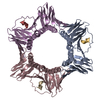
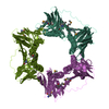
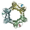
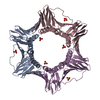
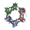
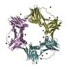
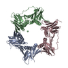
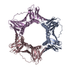
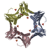
[English] 日本語
Yorodumi
- PDB-6k3a: Crystal structure of human PCNA in complex with DNMT1 PIP box motif. -

+
Open data


ID or keywords:

Loading...

-
Basic information

Entry
Database: PDB / ID: 6k3a
TitleCrystal structure of human PCNA in complex with DNMT1 PIP box motif.
Components
  • Peptide from DNA (cytosine-5)-methyltransferase 1
  • Proliferating cell nuclear antigen
KeywordsREPLICATION / DNMT1 / PCNA / DNA methylation / PIP box
Function / homology
Function and homology information


chromosomal DNA methylation maintenance following DNA replication / negative regulation of vascular associated smooth muscle cell differentiation involved in phenotypic switching / epigenetic programming of gene expression / DNA-methyltransferase activity / negative regulation of vascular associated smooth muscle cell apoptotic process / positive regulation of deoxyribonuclease activity / dinucleotide insertion or deletion binding / PCNA-p21 complex / mitotic telomere maintenance via semi-conservative replication / cellular response to bisphenol A ...chromosomal DNA methylation maintenance following DNA replication / negative regulation of vascular associated smooth muscle cell differentiation involved in phenotypic switching / epigenetic programming of gene expression / DNA-methyltransferase activity / negative regulation of vascular associated smooth muscle cell apoptotic process / positive regulation of deoxyribonuclease activity / dinucleotide insertion or deletion binding / PCNA-p21 complex / mitotic telomere maintenance via semi-conservative replication / cellular response to bisphenol A / DNA (cytosine-5-)-methyltransferase / DNA (cytosine-5-)-methyltransferase activity / purine-specific mismatch base pair DNA N-glycosylase activity / nuclear lamina / positive regulation of DNA-directed DNA polymerase activity / Polymerase switching / MutLalpha complex binding / Telomere C-strand (Lagging Strand) Synthesis / SUMOylation of DNA methylation proteins / Processive synthesis on the lagging strand / PCNA complex / Removal of the Flap Intermediate / female germ cell nucleus / Processive synthesis on the C-strand of the telomere / STAT3 nuclear events downstream of ALK signaling / Polymerase switching on the C-strand of the telomere / Mismatch repair (MMR) directed by MSH2:MSH3 (MutSbeta) / Mismatch repair (MMR) directed by MSH2:MSH6 (MutSalpha) / Removal of the Flap Intermediate from the C-strand / methyl-CpG binding / Transcription of E2F targets under negative control by DREAM complex / replisome / response to L-glutamate / DNA methylation-dependent constitutive heterochromatin formation / negative regulation of gene expression via chromosomal CpG island methylation / response to dexamethasone / histone acetyltransferase binding / lncRNA binding / DNA polymerase processivity factor activity / leading strand elongation / G1/S-Specific Transcription / replication fork processing / nuclear replication fork / pericentric heterochromatin / SUMOylation of DNA replication proteins / PCNA-Dependent Long Patch Base Excision Repair / response to cadmium ion / translesion synthesis / mismatch repair / estrous cycle / cyclin-dependent protein kinase holoenzyme complex / Nuclear events stimulated by ALK signaling in cancer / positive regulation of vascular associated smooth muscle cell proliferation / Translesion synthesis by REV1 / Translesion synthesis by POLK / base-excision repair, gap-filling / DNA polymerase binding / Translesion synthesis by POLI / Gap-filling DNA repair synthesis and ligation in GG-NER / epithelial cell differentiation / liver regeneration / positive regulation of DNA repair / DNA methylation / TP53 Regulates Transcription of Genes Involved in G2 Cell Cycle Arrest / positive regulation of DNA replication / Termination of translesion DNA synthesis / replication fork / Recognition of DNA damage by PCNA-containing replication complex / nuclear estrogen receptor binding / PRC2 methylates histones and DNA / Translesion Synthesis by POLH / Defective pyroptosis / male germ cell nucleus / cellular response to amino acid stimulus / promoter-specific chromatin binding / HDR through Homologous Recombination (HRR) / Dual Incision in GG-NER / NoRC negatively regulates rRNA expression / receptor tyrosine kinase binding / Dual incision in TC-NER / Gap-filling DNA repair synthesis and ligation in TC-NER / cellular response to xenobiotic stimulus / cellular response to hydrogen peroxide / cellular response to UV / E3 ubiquitin ligases ubiquitinate target proteins / response to estradiol / heart development / chromatin organization / methylation / damaged DNA binding / chromosome, telomeric region / nuclear body / negative regulation of gene expression / DNA-templated transcription / centrosome / chromatin binding / positive regulation of gene expression / chromatin / protein-containing complex binding / enzyme binding
Similarity search - Function
DNA (cytosine-5)-methyltransferase 1-like / DNA (cytosine-5)-methyltransferase 1, replication foci domain / Cytosine specific DNA methyltransferase replication foci domain / DMAP1-binding Domain / DMAP1-binding Domain / DMAP1-binding domain / DMAP1-binding domain profile. / : / DNA methylase, C-5 cytosine-specific, conserved site / C-5 cytosine-specific DNA methylases C-terminal signature. ...DNA (cytosine-5)-methyltransferase 1-like / DNA (cytosine-5)-methyltransferase 1, replication foci domain / Cytosine specific DNA methyltransferase replication foci domain / DMAP1-binding Domain / DMAP1-binding Domain / DMAP1-binding domain / DMAP1-binding domain profile. / : / DNA methylase, C-5 cytosine-specific, conserved site / C-5 cytosine-specific DNA methylases C-terminal signature. / Box / Proliferating Cell Nuclear Antigen / Proliferating Cell Nuclear Antigen - #10 / CXXC zinc finger domain / Zinc finger, CXXC-type / Zinc finger CXXC-type profile. / DNA methylase, C-5 cytosine-specific, active site / C-5 cytosine-specific DNA methylases active site. / C-5 cytosine-specific DNA methylase (Dnmt) domain profile. / C-5 cytosine methyltransferase / C-5 cytosine-specific DNA methylase / Bromo adjacent homology domain / BAH domain / Bromo adjacent homology (BAH) domain / Bromo adjacent homology (BAH) domain superfamily / BAH domain profile. / Proliferating cell nuclear antigen signature 2. / Proliferating cell nuclear antigen, PCNA, conserved site / Proliferating cell nuclear antigen signature 1. / Proliferating cell nuclear antigen, PCNA / Proliferating cell nuclear antigen, PCNA, N-terminal / Proliferating cell nuclear antigen, PCNA, C-terminal / Proliferating cell nuclear antigen, N-terminal domain / Proliferating cell nuclear antigen, C-terminal domain / : / S-adenosyl-L-methionine-dependent methyltransferase superfamily / Alpha Beta
Similarity search - Domain/homology
DNA methyltransferase 1 / Proliferating cell nuclear antigen / DNA (cytosine-5)-methyltransferase 1
Similarity search - Component
Biological speciesHomo sapiens (human)
MethodX-RAY DIFFRACTION / SYNCHROTRON / MOLECULAR REPLACEMENT / Resolution: 2.3 Å
AuthorsJimenji, T. / Kori, S. / Arita, K.
Funding support Japan, 2items
OrganizationGrant numberCountry
Ministry of Education, Culture, Sports, Science and Technology (Japan)18H02392 Japan
Japan Science and Technology14530337 Japan
CitationJournal: Biochem.Biophys.Res.Commun. / Year: 2019
Title: Structure of PCNA in complex with DNMT1 PIP box reveals the basis for the molecular mechanism of the interaction.
Authors: Jimenji, T. / Matsumura, R. / Kori, S. / Arita, K.
History
DepositionMay 17, 2019Deposition site: PDBJ / Processing site: PDBJ
Revision 1.0Jun 26, 2019Provider: repository / Type: Initial release
Revision 1.1Jul 10, 2019Group: Data collection / Database references / Category: citation
Item: _citation.country / _citation.journal_abbrev ..._citation.country / _citation.journal_abbrev / _citation.journal_id_ASTM / _citation.journal_id_CSD / _citation.journal_id_ISSN / _citation.pdbx_database_id_DOI / _citation.pdbx_database_id_PubMed / _citation.title / _citation.year
Revision 1.2Jul 24, 2019Group: Data collection / Database references / Category: citation
Item: _citation.journal_volume / _citation.page_first / _citation.page_last
Revision 1.3Nov 22, 2023Group: Data collection / Database references / Refinement description
Category: chem_comp_atom / chem_comp_bond ...chem_comp_atom / chem_comp_bond / database_2 / pdbx_initial_refinement_model
Item: _database_2.pdbx_DOI / _database_2.pdbx_database_accession

-
Structure visualization

Structure viewerMolecule:
MolmilJmol/JSmol

Downloads & links

-
Assembly

Deposited unit
A: Proliferating cell nuclear antigen
B: Peptide from DNA (cytosine-5)-methyltransferase 1
C: Proliferating cell nuclear antigen
D: Peptide from DNA (cytosine-5)-methyltransferase 1
E: Proliferating cell nuclear antigen
F: Peptide from DNA (cytosine-5)-methyltransferase 1


Theoretical massNumber of molelcules
Total (without water)93,6776
Polymers93,6776
Non-polymers00
Water82946
1


  • Idetical with deposited unit
  • defined by author&software
  • Evidence: isothermal titration calorimetry
TypeNameSymmetry operationNumber
identity operation1_555x,y,z1
Buried area8110 Å2
ΔGint-47 kcal/mol
Surface area30950 Å2
MethodPISA
Unit cell
Length a, b, c (Å)141.540, 81.598, 68.105
Angle α, β, γ (deg.)90.00, 90.02, 90.00
Int Tables number5
Space group name H-MC121
Components on special symmetry positions
IDModelComponents
11C-301-

HOH

-
Components

#1: Protein Proliferating cell nuclear antigen / PCNA / Cyclin


Mass: 29006.963 Da / Num. of mol.: 3
Source method: isolated from a genetically manipulated source
Source: (gene. exp.) Homo sapiens (human) / Gene: PCNA / Production host: Escherichia coli (E. coli) / References: UniProt: P12004
#2: Protein/peptide Peptide from DNA (cytosine-5)-methyltransferase 1


Mass: 2218.557 Da / Num. of mol.: 3 / Source method: obtained synthetically / Source: (synth.) Homo sapiens (human) / References: UniProt: K7ERQ1, UniProt: P26358*PLUS
#3: Water ChemComp-HOH / water


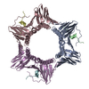
Mass: 18.015 Da / Num. of mol.: 46 / Source method: isolated from a natural source / Formula: H2O

-
Experimental details

-
Experiment

ExperimentMethod: X-RAY DIFFRACTION / Number of used crystals: 1

-
Sample preparation

CrystalDensity Matthews: 2.1 Å3/Da / Density % sol: 41.41 %
Crystal growTemperature: 293 K / Method: vapor diffusion / pH: 6.5 / Details: 1.6M sodium citrate

-
Data collection

DiffractionMean temperature: 100 K / Serial crystal experiment: N
Diffraction sourceSource: SYNCHROTRON / Site: Photon Factory / Beamline: BL-17A / Wavelength: 0.98 Å
DetectorType: DECTRIS PILATUS3 6M / Detector: PIXEL / Date: Dec 15, 2017
RadiationProtocol: SINGLE WAVELENGTH / Monochromatic (M) / Laue (L): M / Scattering type: x-ray
Radiation wavelengthWavelength: 0.98 Å / Relative weight: 1
ReflectionResolution: 2.3→40.84 Å / Num. obs: 34458 / % possible obs: 99.7 % / Redundancy: 3.2 % / CC1/2: 0.997 / Rmerge(I) obs: 0.062 / Net I/av σ(I): 8.2 / Net I/σ(I): 0
Reflection shellResolution: 2.3→2.38 Å / Redundancy: 3.2 % / Rmerge(I) obs: 0.664 / Mean I/σ(I) obs: 1.5 / Num. unique obs: 3366 / CC1/2: 0.705 / % possible all: 99.9

-
Processing

Software
NameVersionClassification
PHENIX(1.15.2_3472: ???)refinement
XDSdata reduction
Aimlessdata scaling
PHASERphasing
RefinementMethod to determine structure: MOLECULAR REPLACEMENT
Starting model: 5MLO
Resolution: 2.3→40.84 Å / SU ML: 0.31 / Cross valid method: FREE R-VALUE / σ(F): 1.33 / Phase error: 30.89
RfactorNum. reflection% reflection
Rfree0.2643 1783 5.19 %
Rwork0.2171 --
obs0.2195 34349 99.35 %
Solvent computationShrinkage radii: 0.9 Å / VDW probe radii: 1.11 Å
Refinement stepCycle: LAST / Resolution: 2.3→40.84 Å
ProteinNucleic acidLigandSolventTotal
Num. atoms5625 0 0 46 5671
Refine LS restraints
Refine-IDTypeDev idealNumber
X-RAY DIFFRACTIONf_bond_d0.0055694
X-RAY DIFFRACTIONf_angle_d0.8527689
X-RAY DIFFRACTIONf_dihedral_angle_d6.3783462
X-RAY DIFFRACTIONf_chiral_restr0.05951
X-RAY DIFFRACTIONf_plane_restr0.005948
LS refinement shell
Resolution (Å)Rfactor RfreeNum. reflection RfreeRfactor RworkNum. reflection RworkRefine-ID% reflection obs (%)
2.3001-2.36220.40191030.31922565X-RAY DIFFRACTION100
2.3622-2.43170.35761600.3012459X-RAY DIFFRACTION100
2.4317-2.51020.32261500.28672437X-RAY DIFFRACTION100
2.5102-2.59990.36811320.2792545X-RAY DIFFRACTION100
2.5999-2.7040.31121360.27212498X-RAY DIFFRACTION100
2.704-2.8270.33031580.27322497X-RAY DIFFRACTION100
2.827-2.9760.35621490.27972473X-RAY DIFFRACTION100
2.976-3.16240.32651340.25892509X-RAY DIFFRACTION99
3.1624-3.40650.36251000.23852547X-RAY DIFFRACTION99
3.4065-3.74910.19741140.20992472X-RAY DIFFRACTION99
3.7491-4.29110.22741590.18142520X-RAY DIFFRACTION99
4.2911-5.40430.21951420.16272511X-RAY DIFFRACTION99
5.4043-40.85050.23061460.20692533X-RAY DIFFRACTION98

+
About Yorodumi

-
News

-
Feb 9, 2022. New format data for meta-information of EMDB entries

New format data for meta-information of EMDB entries

  • Version 3 of the EMDB header file is now the official format.
  • The previous official version 1.9 will be removed from the archive.

Related info.:EMDB header

External links:wwPDB to switch to version 3 of the EMDB data model

-
Aug 12, 2020. Covid-19 info

Covid-19 info

URL: https://pdbj.org/emnavi/covid19.php

New page: Covid-19 featured information page in EM Navigator.

Related info.:Covid-19 info / Mar 5, 2020. Novel coronavirus structure data

+
Mar 5, 2020. Novel coronavirus structure data

Novel coronavirus structure data

Related info.:Yorodumi Speices / Aug 12, 2020. Covid-19 info

External links:COVID-19 featured content - PDBj / Molecule of the Month (242):Coronavirus Proteases

+
Jan 31, 2019. EMDB accession codes are about to change! (news from PDBe EMDB page)

EMDB accession codes are about to change! (news from PDBe EMDB page)

  • The allocation of 4 digits for EMDB accession codes will soon come to an end. Whilst these codes will remain in use, new EMDB accession codes will include an additional digit and will expand incrementally as the available range of codes is exhausted. The current 4-digit format prefixed with “EMD-” (i.e. EMD-XXXX) will advance to a 5-digit format (i.e. EMD-XXXXX), and so on. It is currently estimated that the 4-digit codes will be depleted around Spring 2019, at which point the 5-digit format will come into force.
  • The EM Navigator/Yorodumi systems omit the EMD- prefix.

Related info.:Q: What is EMD? / ID/Accession-code notation in Yorodumi/EM Navigator

External links:EMDB Accession Codes are Changing Soon! / Contact to PDBj

+
Jul 12, 2017. Major update of PDB

Major update of PDB

  • wwPDB released updated PDB data conforming to the new PDBx/mmCIF dictionary.
  • This is a major update changing the version number from 4 to 5, and with Remediation, in which all the entries are updated.
  • In this update, many items about electron microscopy experimental information are reorganized (e.g. em_software).
  • Now, EM Navigator and Yorodumi are based on the updated data.

External links:wwPDB Remediation / Enriched Model Files Conforming to OneDep Data Standards Now Available in the PDB FTP Archive

-
Yorodumi

Thousand views of thousand structures

  • Yorodumi is a browser for structure data from EMDB, PDB, SASBDB, etc.
  • This page is also the successor to EM Navigator detail page, and also detail information page/front-end page for Omokage search.
  • The word "yorodu" (or yorozu) is an old Japanese word meaning "ten thousand". "mi" (miru) is to see.

Related info.:EMDB / PDB / SASBDB / Comparison of 3 databanks / Yorodumi Search / Aug 31, 2016. New EM Navigator & Yorodumi / Yorodumi Papers / Jmol/JSmol / Function and homology information / Changes in new EM Navigator and Yorodumi

Read more